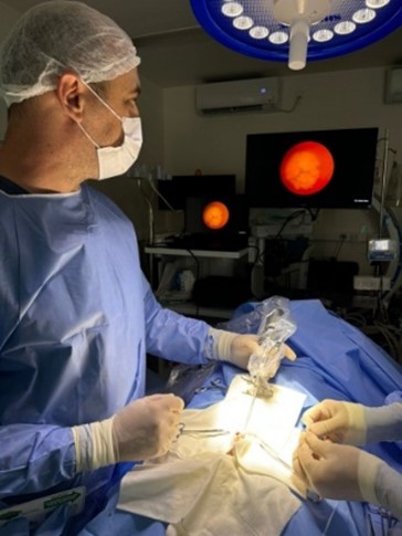
Laparoscopic cholecystectomy in dogs. Biliary sludge in a mixed breed dog
Case report
DOI:
https://doi.org/10.31533/pubvet.v19n01e1713Keywords:
biliary sludge, Cholecystectomy, veterinary medicine, videolaparoscopyAbstract
The development of minimally invasive surgeries has revolutionized the field of surgery. The technique presented in this paper was introduced in human medicine in 1987 and, in recent decades, it has gained importance in veterinary medicine due to its advantages and indications for diagnostic and therapeutic purposes. Videolaparoscopic cholecystectomy is a surgical technique that has come to be used to solve several pathological conditions of the gallbladder, such as biliary sludge, symptomatic cholelithiasis, neoplasms, mucoceles and trauma, especially when these do not respond to drug and clinical treatment with choleretic and lipid-lowering drugs. The procedure consists of small abdominal incisions to remove the gallbladder, with the aid of a laparoscope and specific instruments. It offers a better prognosis when compared to conventional (open) surgery, as the latter is more traumatic and requires longer recovery times. The main advantages of videolaparoscopy are the smaller surgical incisions, reducing surgical stress and postoperative pain, as well as decreased morbidity and rapid recovery, including lower risk of infection and lower incidence of postoperative complications. The present study aims to report the case of a rescued, geriatric, female mixed-breed dog with biliary sludge, as well as to discuss the management of the patient, correlate the laboratory and ultrasound findings with the symptoms, progression and resolution of the treatment using surgical techniques.
References
Agthe, P., Caine, A. R., Posch, B., & Herrtage, M. E. (2009). Ultrasonographic appearance of jejunal lymph nodes in dogs without clinical signs of gastrointestinal disease. Veterinary Radiology & Ultrasound, 50(2), 195–200. https://doi.org/10.1111/j.1740-8261.2009.01516.x.
Aguirre, A. L., Center, S. A., Randolph, J. E., Yeager, A. E., Keegan, A. M., Harvey, H. J., & Erb, H. N. (2007). Gallbladder disease in Shetland Sheepdogs: 38 Cases (1995-2005). Journal of the American Veterinary Medical Association, 231(1), 79–88. https://doi.org/10.2460/javma.231.1.79.
Amboldi, M., Amboldi, A., Gherardi, G., & Bonandrini, L. (2011). Complications of videolaparoscopic cholecystectomy: A retrospective analysis of 1037 consecutive cases. International Surgery, 96(1), 35–44. https://doi.org/10.9738/1385.1.
Bakulin, I. G., Avalueva, E. B., Serkova, M. U., Skvortsova, T. E., Seliverstov, P. V., Shevyakov, M. A., & Sitkin, S. I. (2021). Biliary sludge: Pathogenesis, etiology and drug therapy. Terapevticheskii Arkhiv, 93(2). https://doi.org/10.26442/00403660.2021.02.200638,
Besso, J. G., Wrigley, R. H., Gliatto, J. M., & Webster, C. R. L. (2000). Ultrasonographic appearance and clinical findings in 14 dogs with gallbladder mucocele. Veterinary Radiology and Ultrasound, 41(3), 261–271. https://doi.org/10.1111/j.1740-8261.2000.tb01489.x,
Brömel, C., Barthez, P. Y., Léveillé, R., & Scrivani, P. V. (1998). Prevalence of gallbladder sludge in dogs as assessed by ultrasonography. Veterinary Radiology and Ultrasound, 39(3), 206–210. https://doi.org/10.1111/j.1740-8261.1998.tb00341.x.
Carrilho, F. J., Mattos, A. A., Vianey, A. F., Vezozzo, D. C. P., Marinho, F., Souto, F. J., Cotrim, H. P., Coelho, H. S. M., Silva, I., Garcia, J. H. P., Kikuchi, L., Lofego, P., Andraus, W., Strauss, E., Silva, G., Altikes, I., Medeiros, J. E., Bittencourt, P. L., & Parise, E. R. (2015). Brazilian society of hepatology recommendations for the diagnosis and treatment of hepatocellular carcinoma. Arquivos de Gastroenterologia, 52(suppl 1). https://doi.org/10.1590/s0004-28032015000500001.
Carvalho, C. F. (2018). Ultrassonografia em pequenos animais. Editora Roca.
Center, S. A. (2009). Diseases of the gallbladder and biliary tree. Veterinary Clinics of North America: Small Animal Practice, 39(3), 543–598.
De Marco, V. (2015). Hiperadrenocorticismo canino. In M. Jericó, J. Andrade Neto, & M. Kogika (Eds.), Tratado de medicina interna de cães e gatos (pp. 1691–1703).
Espíndola, R. F. (2014). Ultrassonografia intervencionista em pequenos animais. Universidade de Brasília.
Ferguson, J. F. (1996). What is your diagnosis? [Calcinosis circumscripta in a dog]. In Generic.
Fossum, T. W. (2021). Cirurgia de pequenos animais (3ed.). Elsevier Editora.
Gaschen, L. (2013). Ultrassonographic Imaging of the gastrointestinal tract. In R. J. Washabau & M. J. Day (Eds.), Diagnostic Imaging of gastrointestinal tract (pp. 228–235). Elsevier.
Hayward, N. (2012). BSAVA Manual of canine and feline ultrasonography. Journal of Small Animal Practice, 53(2). https://doi.org/10.1111/j.1748-5827.2012.01183.x.
Herrtage, M. E., & Ramsey, I. K. (2015). Hiperadrenocorticismo em cães. In C. T. Mooney & M. E. Peterson (Eds.), Manual de Endocrinologia em Cães e Gatos (Vol. 4, pp. 254–289). Koog.
Jaffey, J. A., Matheson, J., Shumway, K., Pacholec, C., Ullal, T., Van Den Bossche, L., Fieten, H., Ringold, R., Lee, K. J., & DeClue, A. E. (2020). Serum 25-hydroxyvitamin D concentrations in dogs with gallbladder mucocele. PLoS ONE, 15(12), e058411. https://doi.org/10.1371/journal.pone.0244102.
Jüngst, C., Kullak-Ublick, G. A., & Jüngst, D. (2006). Microlithiasis and sludge. Best Practice and Research: Clinical Gastroenterology, 20(6). https://doi.org/10.1016/j.bpg.2006.03.007.
Junqueira, L. C., & Carneiro, J. C. (2013). Histologia Básica (12 ed.). Guanabara Koogan.
Klein, B. G. (2014). Cunningham Tratado de Fisiologia Veterinária. Elsevier.
Klinkspoor, J. H., Kuver, R., Savard, C. E., Oda, D., Azzouz, H., Tytgat, G. N. J., Groen, A. K., & Lee, S. P. (1995). Model bile and bile salts accelerate mucin secretion by cultured dog gallbladder epithelial cells. Gastroenterology, 109(1), 264–274. https://doi.org/10.1016/0016-5085(95)90293-7.
Malek, S., Sinclair, E., Hosgood, G., Moens, N. M. M., Baily, T., & Boston, S. E. (2013). Clinical findings and prognostic factors for dogs undergoing cholecystectomy for gall bladder mucocele. Veterinary Surgery, 42(4), 418–426. https://doi.org/10.1111/j.1532-950X.2012.01072.x.
Mayhew, P. D. (2009). Advanced laparoscopic procedures (hepatobiliary, endocrine) in dogs and cats. In Veterinary Clinics of North America - Small Animal Practice (Vol. 39, Issue 5, pp. 925–939). https://doi.org/10.1016/j.cvsm.2009.05.004,
Mizutani, S., Torisu, S., Kaneko, Y., Yamamoto, S., Fujimoto, S., Ong, B. H. E., & Naganobu, K. (2017). Retrospective analysis of canine gallbladder contents in biliary sludge and gallbladder mucoceles. Journal of Veterinary Medical Science, 79(2). https://doi.org/10.1292/jvms.16-0562.
Nagao, I., Tsuji, K., Goto-Koshino, Y., Tsuboi, M., Chambers, J. K., Uchida, K., Kambayashi, S., Tomiyasu, H., Baba, K., & Okuda, M. (2023). MUC5AC and MUC5B expression in canine gallbladder mucocele epithelial cells. Journal of Veterinary Medical Science, 85(12), 1269–1276. https://doi.org/10.1292/jvms.23-0174.
Nyland, T. G., & Mattoon, J. S. (2005). Ultra-som diagnóstico em pequenos animais. Editora Roca.
Parkanzky, M., Grimes, J., Schmiedt, C., Secrest, S., & Bugbee, A. (2019). Long-term survival of dogs treated for gallbladder mucocele by cholecystectomy, medical management, or both. Journal of Veterinary Internal Medicine, 33(5), 10–17. https://doi.org/10.1111/jvim.15611.
Pazzi, P., Gamberini, S., Buldrini, P., & Gullini, S. (2003). Biliary sludge: The sluggish gallbladder. Digestive and Liver Disease, 35(SUPPL. 3), 19–45. https://doi.org/10.1016/S1590-8658(03)00093-8.
Penninck, D. G., & D’Anjou, M. A. (2011). Atlas de ultrassonografia de Pequenos animais (p. 513p.). Guanabara Koogan.
Piñol, F., Ruiz, J., Segura, N., Proaño, P., & Sánchez, E. (2020). La vesícula biliar como reservorio y protectora del tracto digestivo. Revista Cubana de Investigaciones Biomédicas, 39(1).
Quinn, P. J. (1994). Clinical veterinary microbiology (Issue SF 780.2. C54 1994).
Quinn, R., & Cook, A. K. (2009). update on gallbladder mucoceles in dogs. Veterinary Medicine, 103(4), 169–175.
Reed, W. H., & Ramirez, S. (2007). What is your diagnosis? Gallbladder mucocele. Journal of the Veterinary Medical Association, 230, 661–662.
Richter, K. P. (2005). Doenças do fígado e do sistema hepatobiliar. In T. R. Tams (Ed.), Gastroenterologia de pequenos animais (pp. 283–348). Roca.
Romero, G. M., Ortuño, L. E. G., Casas, F. C., Carvajal, K. S., & Aguilar, R. E. M. (2008). Mucocele en la vesícula biliar de un perro: hallazgos clínico-patológicos. Veterinária Mexico, 39(3), 335–340.
Salim, M. T., & Cutait, R. (2008). Complicações da cirurgia videolaparoscópica no tratamento de doenças da vesícula e vias biliares. ABCD. Arquivos Brasileiros de Cirurgia Digestiva (São Paulo), 21(4), 153–157. https://doi.org/10.1590/s0102-67202008000400001.
Saucedo, L. G. R., & Martins, W. P. (2009). Ultra-sonografia endoscópica nos quadros de pancreatite. Experts in Ultrasound: Reviews and Perspectives, 1(2), 113–124. https://doi.org/10.4281/eurp.2009.02.09,
Seoane, M. P. R., Garcia, D. A. A., & Froes, T. R. (2011). A história da ultrassonografia veterinária em pequenos animais. Archives of Veterinary Science, 16(1), 54–61.
Silva, F. C. K., Drumond, J. P., & Coelho, N. G. D. (2022). Hiperadrenocorticismo canino: Revisão. PUBVET, 16(5), 1–7. https://doi.org/10.31533/pubvet.v16n05a1125.1-7.
Tsukagoshi, T., Ohno, K., Tsukamoto, A., Fukushima, K., Takahashi, M., Nakashima, K., Fujino, Y., & Tsujimoto, H. (2012). Decreased gallbladder emptying in dogs with biliary sludge or gallbladder mucocele. Veterinary Radiology & Ultrasound, 53(1), 84–91. https://doi.org/10.1111/j.1740-8261.2011.01868.x.
Walter, R., Dunn, M. E., D’Anjou, M. A., & Lécuyer, M. (2008). Nonsurgical resolution of gallbladder mucocele in two dogs. Journal of the American Veterinary Medical Association, 232(11), 1688–1693. https://doi.org/10.2460/javma.232.11.1688.
Zavaleta-García, L. del C., & Ortiz-Hidalgo, C. (2023). La vesícula biliar: Un recorrido microscópico por su anatomía normal y algunas implicaciones patológicas. Patología Revista Latinoamericana.
Zorniak, M., Sirtl, S., Beyer, G., Mahajan, U. M., Bretthauer, K., Schirra, J., Schulz, C., Kohlmann, T., Lerch, M. M., & Mayerle, J. (2023). Consensus definition of sludge and microlithiasis as a possible cause of pancreatitis. Gut, 72(10). https://doi.org/10.1136/gutjnl-2022-327955.

Downloads
Published
Issue
Section
License
Copyright (c) 2024 Débora Todeschini de Castro, Edelmar Chagas da Silva, Ingrid da Silveira Chiarelli, Mariana Tavares Maioni, Professora Msc. Patrícia Franciscone Mendes, M.V. André Preturlon Terra

This work is licensed under a Creative Commons Attribution 4.0 International License.
Você tem o direito de:
Compartilhar — copiar e redistribuir o material em qualquer suporte ou formato
Adaptar — remixar, transformar, e criar a partir do material para qualquer fim, mesmo que comercial.
O licenciante não pode revogar estes direitos desde que você respeite os termos da licença. De acordo com os termos seguintes:
Atribuição
— Você deve dar o crédito apropriado, prover um link para a licença e indicar se mudanças foram feitas. Você deve fazê-lo em qualquer circunstância razoável, mas de nenhuma maneira que sugira que o licenciante apoia você ou o seu uso. Sem restrições adicionais
— Você não pode aplicar termos jurídicos ou medidas de caráter tecnológico que restrinjam legalmente outros de fazerem algo que a licença permita.


