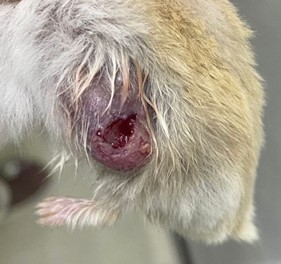
Cutaneous hemangiossarcoma in Phodopus roboroviskii
DOI:
https://doi.org/10.31533/pubvet.v19n01e1716Keywords:
Cutaneous hemangiosarcoma, hamster, neoplasm, rodentAbstract
This work describes a clinical case of cutaneous hemangiosarcoma in a Roborovski hamster (Phodopus roborovskii). There are few reports of neoplasms in hamsters of this species, making this the first record of cutaneous hemangiosarcoma, a malignant mesenchymal neoplasm characterized by autonomous and invasive cell growth with metastatic potential. The diagnosis of hemangiosarcoma was made through clinical examination, radiography, and biopsy of a subcutaneous nodule located on the left flank of the animal. The male hamster, approximately 3 years old, presented with an ulcerated nodule which was subjected to surgical excision for histopathological analysis, confirming the diagnosis of cutaneous hemangiosarcoma. The surgical procedure was successfully performed, with the animal under inhalational anesthesia using sevoflurane at a dose of 1.5 MAC and local anesthesia with lidocaine at a dose of 2 mg/kg. Post-operative treatment included the administration of analgesics and anti-inflammatories, with continuous clinical follow-up. Histopathological analysis revealed a neoplasm composed of endothelial cells with moderate pleomorphism, areas of hemorrhage, fibroplasia, and inflammatory infiltration. No signs of metastasis were observed in the pre- and post-operative radiographs. This study highlights the importance of early diagnosis and surgical intervention in small rodents, as well as it marks the first report of cutaneous hemangiosarcoma in Roborovskii hamsters according to the consulted scientific literature, and underscores the need for further research to better understand neoplasms in this species.
References
Dutton, M. (2020). Selected veterinary concerns of geriatric rats, mice, hamsters, and gerbils. Veterinary Clinics: Exotic Animal Practice, 23(3), 525–548. https://doi.org/10.1016/j.cvex.2020.04.001.
Fernandes, S. C., & Nardi, A. D. B. N. (2016). Hemangiossarcomas. In C. R. Daleck, A. B. De Narde, & S. Rodaski (Eds.), Oncologia em cães e gatos (pp. 776–796). Roca, Brasil.
Greaves, P., Chouinard, L., Ernst, H., Mecklenburg, L., Pruimboom-brees, I. M., Rinke, M., Rittinghausen, S., Thibault, S., Erichsen, J. von, & Yoshida, T. (2013). Proliferative and Non-Proliferative Lesions of the Rat and Mouse Soft Tissue, Skeletal Muscle and Mesothelium. Journal of Toxicologic Pathology, 26(3_Suppl). https://doi.org/10.1293/tox.26.1s.
Hsieh, C. Y., Tsai, H. W., Chang, C. C., Lin, T. W., Chang, K. C., & Chen, Y. S. (2015). Tumors involving skin, soft tissue and skeletal muscle: Benign, primary malignant or metastatic? Asian Pacific Journal of Cancer Prevention, 16(15). https://doi.org/10.7314/APJCP.2015.16.15.6681.
Kiehl, A. R., & Mays, M. B. C. (2016). Atlas for the Diagnosis of Tumors in the Dog and Cat. In Atlas for the Diagnosis of Tumors in the Dog and Cat. https://doi.org/10.1002/9781119050766.
Kling, M. A. (2011). A Review of Respiratory System Anatomy, Physiology, and Disease in the Mouse, Rat, Hamster, and Gerbil. In Veterinary Clinics of North America - Exotic Animal Practice (Vol. 14, Issue 2). https://doi.org/10.1016/j.cvex.2011.03.007.
Kondo, H., Onuma, M., Shibuya, H., & Sato, T. (2008). Spontaneous tumors in domestic hamsters. Veterinary Pathology, 45(5), 674–680. https://doi.org/10.1354/vp.45-5-674.
Kubiak, M. (2020). Handbook of Exotic Pet Medicine. In Handbook of Exotic Pet Medicine. https://doi.org/10.1002/9781119389934
Machado, P. C., Salzedas, B. A., Segala, R. D., & Pita, M. C. G. (2021). Hemangiossarcoma em hamster sírio (Mesocricetus auratus) – Relato de caso. Brazilian Journal of Animal and Environmental Research, 4(1), 1134–1147. https://doi.org/10.34188/bjaerv4n1-090.
Megha, K. B., Joseph, X., Akhil, V., & Mohanan, P. V. (2021). Cascade of immune mechanism and consequences of inflammatory disorders. In Phytomedicine (Vol. 91). https://doi.org/10.1016/j.phymed.2021.153712.
Meredith, A., & Redrobe, S. (2002). BSAVA Manual of Exotic Pets. BSAVA Manual of Exotic Pets.
Parkinson, L. (2023). Fluid Therapy in Exotic Animal Emergency and Critical Care. In Veterinary Clinics of North America - Exotic Animal Practice (Vol. 26, Issue 3). https://doi.org/10.1016/j.cvex.2023.05.004.
Pereira, R. M. F., Lima, T. S., Oliveira, R. L., Fonseca, S. M. C., Wicpolt, N. S., Farias, R. C., Lucena, R. B., Pavarini, S. P., Araújo, J. L. de, & Mendonça, F. S. (2024). Clinical, pathological and immunohistochemical characterization of spontaneous neoplasms in pet rodents in Northeastern Brazil. Pesquisa Veterinaria Brasileira, 44. https://doi.org/10.1590/1678-5150-PVB-7410.
Quesenberry, K., & Carpenter, J. W. (2011). Ferrets, rabbits and rodents: clinical medicine and surgery. Elsevier Health Sciences.
Rother, N., Bertram, C. A., Klopfleisch, R., Fragoso-Garcia, M., Bomhard, W. V., Schandelmaier, C., & Müller, K. (2021). Tumours in 177 pet hamsters. Veterinary Record, 188(6). https://doi.org/10.1002/vetr.14.
Sayers, I., & Smith, S. (2010). Mice, rats, hamsters and gerbils. In A. Meredith & C. Johnson-Delaney (Eds.), BSVA Manual of exotic pets (pp. 1–27). https://doi.org/10.22233/9781905319909.1.
Schultheiss, P. C. (2004). A retrospective study of visceral and nonvisceral hemangiosarcoma and hemangiomas in domestic animals. Journal of Veterinary Diagnostic Investigation, 16(6), 522–526. https://doi.org/10.1177/104063870401600606.
Sharkey, L. C., Seelig, D. M., & Overmann, J. (2014). All lesions great and small, part 1: diagnostic cytology in veterinary medicine. Diagnostic Cytopathology, 42(6), 535–543. https://doi.org/10.1002/dc.23097.
Sharun, K., Basha, M. A., Shah, M. A., Kumar, K., Kumar, P., Shivaraju, S., Pawde, A. M., & Amarpal. (2019). Clinical management of cutaneous hemangiosarcoma in canines: a review of five cases. Comparative Clinical Pathology, 28(6). https://doi.org/10.1007/s00580-019-03039-1.
Sulzbach, S. F., Tanaka, G. A., & Rumpel, A. S. (2023). A domesticação e sua influência na criação e bem-estar de hamsters: Revisão. PUBVET, 17(3), 1–6. https://doi.org/10.31533/pubvet.v17n03a1358.
Tizziani Júnior, E., Oliveira, G. M. D., & Souza, M. T. (2023). Exérese de um hemangiossarcoma cutâneo facial: Relato de caso. PUBVET, 17(5), e1381. https://doi.org/10.31533/pubvet.v17n5e1381.

Downloads
Published
Issue
Section
License
Copyright (c) 2024 Ana Carolyne Borges de Oliveira, Karla Vitória Alves Sampaio, Isabella Saad Martins da Silva, Gabriela Scarpin de Souza, Marcus Vinícius Lima David, Danielle Nascimento Silva, Paulo Roberto Bahiano Ferreira

This work is licensed under a Creative Commons Attribution 4.0 International License.
Você tem o direito de:
Compartilhar — copiar e redistribuir o material em qualquer suporte ou formato
Adaptar — remixar, transformar, e criar a partir do material para qualquer fim, mesmo que comercial.
O licenciante não pode revogar estes direitos desde que você respeite os termos da licença. De acordo com os termos seguintes:
Atribuição
— Você deve dar o crédito apropriado, prover um link para a licença e indicar se mudanças foram feitas. Você deve fazê-lo em qualquer circunstância razoável, mas de nenhuma maneira que sugira que o licenciante apoia você ou o seu uso. Sem restrições adicionais
— Você não pode aplicar termos jurídicos ou medidas de caráter tecnológico que restrinjam legalmente outros de fazerem algo que a licença permita.


