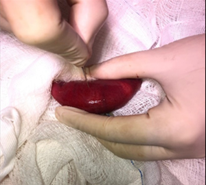
Enterotomy to remove a foreign body in the jejunum
DOI:
https://doi.org/10.31533/pubvet.v18n11e1683Keywords:
Emesis, imaging tests, laparotomy, obstructionAbstract
In the small animal clinic, dogs are frequently seen that ingest non-digestible objects which end up causing obstruction in the animal's digestive system. Young canines are the most affected, as they tend to eat or/and play with inedible objects indiscriminately. The diagnosis of intestinal obstruction is essential, as it can cause clinical repercussions such as erosions and ulcers. Therefore, it is based on anamnesis and clinical signs such as emesis, anorexia and apathy. In addition, when necessary, the professional may request imaging tests and an exploratory laparotomy to confirm the diagnostic hypothesis. From this perspective, a Shih Tzu dog, approximately two years old, weighing 4 kg, was seen at the clinic. During anamnesis, the guardian reports that the patient was apathetic and had recurrent vomiting. Upon clinical examination, only moderate dehydration was evident. The treatment was carried out surgically using the laparotomy technique followed by enterotomy, which is the most recommended treatment in cases of intestinal obstruction caused by a foreign body. The animal had a positive prognosis and a good recovery. Therefore, the aim of this work is to report a case of obstruction of the intestinal tract by a foreign body in a dog, and the surgical techniques used to treat it.
References
Assunção, G. A. (2017). Corpos estranhos esofágicos em cães e gatos: Revisão de literatura. Universidade Federal do Rio Grande do Sul.
Bernardo, R. F. B., Varallo, G. R., & Silveira, R. N. (2023). Conduta diagnóstica e terapêutica para corpo estranho linear em gato: Relato de caso. PUBVET, 17(1), 1–6. https://doi.org/10.31533/pubvet.v17n01a1334.
Bojrab, M. J. (2014). Mecanismos das doenças em cirurgia de pequenos animais (Vol. 1). Roca.
Bongard, A. B., Furrow, E., & Granick, J. L. (2019). Retrospective evaluation of factors associated with degree of esophagitis, treatment, and outcomes in dogs presenting with esophageal foreign bodies (2004–2014): 114 cases. Journal of Veterinary Emergency and Critical Care, 29(5), 528–534. https://doi.org/10.1111/vec.12875.
Castro, J. S., Menezes, M. T., Rossignoli, P. P. R., Gondim, B. S., Reis, V. R., & Pieroni, P. M. R. L. (2024). Corpo estranho linear intestinal em cão: Relato de caso. PUBVET, 17(13). https://doi.org/10.31533/pubvet.v17n13e1519.
Fantoni, D. T., & Cortopassi, S. R. G. (2009). Anestesia em cães e gatos. Roca.
Fossum, T. W. (2021). Cirurgia de pequenos animais (3ed.). Elsevier Editora.
Gupta, P., Sharma, A., Gupta, A. K., Kushwaha, R. B., & Dwivedi, D. K. (2012). Gastric foreign body and its surgical management in a dog. Intas, 13(1).
Jericó, M. M., Andrade Neto, J. P., & Kogika, M. M. (2015). Tratado de medicina interna de cães e gatos. Roca Ltda.
Lumb, W. V, Jones, E. W., Téllez, E., & Retana, R. (2017). Anestesia veterinária. Continental.
Macphail, C. M. (2013). Corpo estranho e obstrução gastrintestinais. In E. M. Mazzaferro (Ed.), Emergências e cuidados críticos em pequenos animais (pp. 131–137). Roca Ltda.
Martinez-Taboada, F., & Leece, E. A. (2014). Comparison of propofol with ketofol, a propofol-ketamine admixture, for induction of anaesthesia in healthy dogs. Veterinary Anaesthesia and Analgesia, 41(6). https://doi.org/10.1111/vaa.12171.
Nelson, R., & Couto, C. G. (2015). Medicina interna de pequenos animais (3.ed.). Elsevier Brasil.
Oliveira, A. L. (2022). Cirurgia veterinária em pequenos animais. Manole, São Paulo, Brasil.
Pahim, A. B. S., Pahim, A. B. S., Valério, G. B., Guim, T. N., Orlandin, R., Martins, F. P., & Oliveira, M. T. (2020). Protocolos de medicação pré-anestésica utilizados no Huvet Unipampa. Anais Do Salão Internacional de Ensino, Pesquisa e Extensão, 12(2).
Parra, T. C., Berno, M. D. B., Guimarães, A., Andrade, L. C. A., Mosquini, A. F., & Montanha, F. P. (2012). Ingestão de corpo estranho em cães–Relato de caso. Revista Científica Eletrônica de Medicina Veterinária, 18.
Silva, F. F. S., Ré, B. G., Pinto, A. C. B. C. F., Lorigados, C. A. B., Unruh, S. M., & Kanayama, L. M. (2016). Diagnóstico por imagem de corpo estranho gastrointestinal em cães e gatos: estudo retrospectivo de 157 casos. Revista de Educação Continuada em Medicina Veterinária e Zootecnia do CRMV-SP, 14(3), 54–55.
Slatter, D. H., & Aronson, L. (2007). Manual de cirurgia de pequenos animais (Vol. 2). Manole São Paulo.
Sousa, E. J. N., Castro, R. J. S., Oliveira, F. A. S., Ferreira, N. L., Cabral, C. F., Fonteles, A. J. S., Silva, P. O., Costa, M. S., & Pereira Júnior, J. R. P. (2022). Avaliação eletrocardiográfica de cães submetidos à medicação pré-anestésica com acepromazina/meperidina ou acepromazina/metadona. PUBVET, 16(3), 1–6. https://doi.org/10.31533/pubvet.v16n03a1056.1-6.
Viana, E. G., Bezerra, S. T. C. S., Rodrigues, I. R., Braga, C. C. S., & Pinto, R. N. (2020). Abordagem clinico-cirúrgica em cão com corpo estranho linear extenso. Ciência Animal, 30(2), 42–50.

Downloads
Published
Issue
Section
License
Copyright (c) 2024 Elyfelette Souza Ferreira, Fernanda Nascimento de Souza, Frankcijhonatan Vasconcelos Camelo Pereira, Roberto Barbuio

This work is licensed under a Creative Commons Attribution 4.0 International License.
Você tem o direito de:
Compartilhar — copiar e redistribuir o material em qualquer suporte ou formato
Adaptar — remixar, transformar, e criar a partir do material para qualquer fim, mesmo que comercial.
O licenciante não pode revogar estes direitos desde que você respeite os termos da licença. De acordo com os termos seguintes:
Atribuição
— Você deve dar o crédito apropriado, prover um link para a licença e indicar se mudanças foram feitas. Você deve fazê-lo em qualquer circunstância razoável, mas de nenhuma maneira que sugira que o licenciante apoia você ou o seu uso. Sem restrições adicionais
— Você não pode aplicar termos jurídicos ou medidas de caráter tecnológico que restrinjam legalmente outros de fazerem algo que a licença permita.


