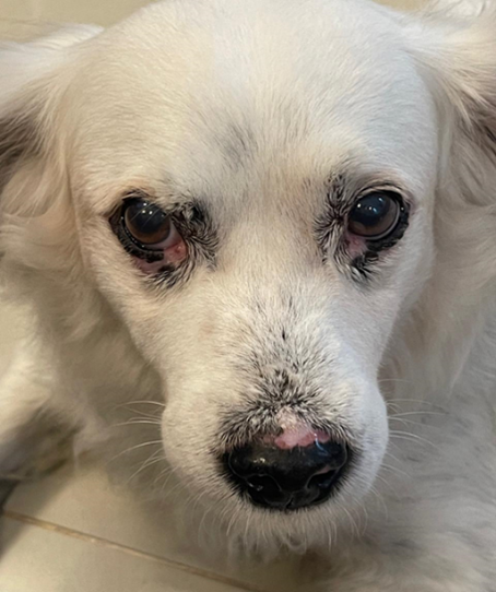
Discoid Lupus Erythematosus in a mixed breed dog
Case report
DOI:
https://doi.org/10.31533/pubvet.v18n09e1657Keywords:
Autoimmune, canine, dermatopathy, depigmentation, ulcerationAbstract
The objective of this work was to report a case of Discoid Lupus Erythematosus (DLE) in a mixed breed dog, 9 years old and weighing 18 kg. Autoimmune cutaneous diseases are rare dermatoses, characterized by the production of autoantibodies that damage mucocutaneous tissue, causing inflammation and tissue damage, with DLE being the second most prevalent in dogs and considered a benign variant of systemic lupus erythematosus. Its etiology is still unknown, but genetic, endocrine and infectious factors, exposure to ultraviolet light and drug reactions are involved. This pathology has already been observed in dogs, cats, horses and humans. The lesional condition consists of depigmentation, eroded-crusted and ulcerative lesions, erythema, alopecia and pruritus, mainly affecting the periocular region, mirror and nasal bridge. Excluding other skin diseases with similar clinical signs and performing a skin biopsy, and subsequent histopathological examination, allow for confirmation of the diagnosis. Therapy for this disease involves the use of sunscreen, immunosuppressive medication and supplementation of fatty acids and vitamins A and E. The patient in this report was initially seen on April 9th, 2021, in the city of Nova Iguaçu (Rio de Janeiro) presenting itching, eroded crusty lesions, erythema and alopecia in the nasal bridge and periocular region, in addition to edema and depigmentation in the nasal mirror, and the same had already been seen in 2018 by another professional, at a different location, who did not request additional exams. However, they did prescribe topical treatment with Crema 6A® ointment, without a positive outcome. The diagnosis was obtained through histopathology of the skin lesions (nasal and labial), which confirmed the DLE. On July 28th, 2023, this patient was seen by another professional at a different veterinary clinic, and PCR (Polymerase Chain Reaction) of skin fragments and serology (ELISA + RIFI with total dilution) for leishmaniasis were performed for differential diagnosis, which yielded negative results. The patient underwent different treatment protocols that were not satisfactory, except for the last (current) one, consisting of immunosuppressive drugs: oral Cyclosporine, and Tacrolimus ointment for topical use in lesions, in addition to the use of sunscreen on the face. The patient in the present study demonstrated a good response, as there was remission of the lesions, erythema and pigmentation, in addition to not presenting any adverse drug reactions.
References
Ataíde, W., Silva, V., Ferraz, H., Amaral, A., & Romani, A. (2019). Lúpus eritematoso discóoide em cães. Enciclopédia Biosfera, 16(29), 1–15. https://doi.org/10.18677/encibio_2019a81.
Banovic, F. (2019). Canine cutaneous lupus erythematosus: Newly discovered variants. Veterinary Clinics: Small Animal Practice, 49(1), 37–45. https://doi.org/10.1016/j.cvsm.2018.08.004.
Bressan, A. L., Silva, S. R. D., Fontenelle, E., & Gripp, A. C. (2010). Agentes imunossupressores em dermatologia. Anais Brasileiros de Dermatologia, 9–22.
Conceição, L. G., & Santos, L. S. (2010). Sistema tegumentar. In L. G. Conceição (Ed.), Patologia Veterinária (Vol. 1, pp. 423–524). Roca.
Ettinger, S. J., Feldman, E. C., & Cote, E. (2017). Textbook of Veterinary Internal Medicine-eBook. Elsevier Health Sciences.
Fragoso, T. S., Dantas, A. T., Marques, C. D. L., Rocha Júnior, L. F., Melo, J. H. L., Costa, A. J. G., & Duarte, A. L. B. P. (2012). Níveis séricos de 25-hidroxivitamina D3 e sua associação com parâmetros clínicos e laboratoriais em pacientes com lupus eritematoso sistêmico. Revista Brasileira de Reumatologia, 52, 60–65. https://doi.org/10.1590/s0482-50042012000100007.
Gorman, N. T. Imunologia. In: Ettinger S.J.; Feldman E.C (1997). Tratado de medicina interna veterinária. 4 ed. São Paulo: Manole 2735 - 2765p.
Guaguère, E., & Bensignor, E. (2005). Terapêutica dermatológica do cão (Vol. 1). Roca.
Guilherme, A. R. C. & Lupp, M. M. C. P. (2024). Diagnóstico de lúpus eritematoso discóide em cão: Relato de caso, PUBVET, 18(8), 1–6. https://doi.org/10.31533/pubvet.v18n08e1645.
Guimarães, F. C., Conceição, R. T., Flaiban, K. K. M. C., & Arias, M. V. B. (2022). Estudo retrospectivo em 18 cães com lúpus eritematoso sistêmico (2008–2018). PUBVET, 16(2), 1–8. https://doi.org/10.31533/pubvet.v16n02a1032.1-8.
Hnilica, K. A., & Patterson, A. P. (2011). Small animal dermatology: a color atlas and therapeutic guide. Elsevier Health Sciences.
Hnilica, K. A., & Patterson, A. P. (2017). Autoimmune and immune-mediated skin disorders. In K. A. Hnilica & P. A.P. (Eds.), Small animal dermatology: a color atlas and therapeutic guide. Elsevier.
Johnson, K. A., Watson, A. D. J., Ettinger, S. J., & Feldman, E. C. (2004). Tratado de Medicina Interna Veterinária: doenças do cão e do gato. Manole Ltda.
Larsson, C. E., & Otsuka, M. (2000). Lúpus Eritematoso Discóide – LED: Revisão e casuística em serviço especializado da capital de São Paulo. Revista de Educação Continuada Em Medicina Veterinária e Zootecnia Do CRMV-SP, 3(1), 29–36. https://doi.org/10.36440/recmvz.v3i1.3349.
Lima-Verde, J. F., Ferreira, T. C., & Nunes-Pinheiro, D. C. S. (2020). Lupus eritematoso discoide em cão: relato de caso. PUBVET, 14(1), 1–6. https://doi.org/10.31533/pubvet.v14n1a486.1-6
MacPhail, C. M. (2014). Cirurgia do sistema tegumentar. In Cirurgia de pequenos animais. Elsevier Rio de Janeiro.
Madan, V., & Griffiths, C. E. M. (2007). Systemic ciclosporin and tacrolimus in dermatology. In Dermatologic Therapy (Vol. 20, Issue 4, pp. 239–250). https://doi.org/10.1111/j.1529-8019.2007.00137.x.
Medleau, L., & Hnilica, K. A. (2006). Small animal dermatology. Editora Roca.
Oberkirchner, U., Linder, K. E., & Olivry, T. (2012). Successful treatment of a novel generalized variant of canine discoid lupus erythematosus with oral hydroxychloroquine. Veterinary Dermatology, 23(1), 65e16. https://doi.org/10.1111/j.1365-3164.2011.00994.x.
Palumbo, M. I. P., Machado, L. H. A., Conti, J. P., Oliveira, F. C., & Rodrigues, J. C. (2010). Incidência das dermatopatias autoimunes em cães e gatos e estudo retrospectivo de 40 casos de lupus eritematoso discóide atendidos no serviço de dermatologia da Faculdade de Medicina Veterinária e Zootecnia da UNESP – Botucatu. Semina: Ciências Agrárias, 31(3), 739–744. https://doi.org/10.5433/1679-0359.2010v31n3p739.
Patel, A., & Forsythe, P. J. (2011). Dermatologia em pequenos animais. Elsevier Brasil.
Rhodes, K. H., & Werner, A. H. (2014). Dermatologia em Pequenos Animais, 2a edição (2 Ed.). Roca, São Paulo.
Santos, R. D. L., & Alessi, A. C. (2017). Sistema Tegumentar. In Patologia Veterinária 471-472. São Paulo, SP: Editora Roca.
Salviatto, C. M. (2021). Lúpus eritematoso discóide: Relato de caso. https://doi.org/10.54265/mrve9681.
Tizard, I. R. (2017). Veterinary Immunology-E-Book. Elsevier Health Sciences.
Tzellos, T. G., & Kouvelas, D. (2008). Topical tacrolimus and pimecrolimus in the treatment of cutaneous lupus erythematosus: An evidence-based 37. In European Journal of Clinical Pharmacology (Vol. 64, Issue 4, pp. 337–341). https://doi.org/10.1007/s00228-007-0421-2.
Vettorato, E. D., & Novais-Mencalha, R. (2023). Lúpus eritematoso cutâneo crônico–Relato de caso. PUBVET, 17(6), e1396. https://doi.org/10.31533/pubvet.v17n6e1396.
Wilkinson, G. T., Harvey, R. G., & Nascimento, F. G. D. (1997). Atlas colorido de dermatologia de pequenos animais: Guia para o diagnóstico.

Downloads
Published
Issue
Section
License
Copyright (c) 2024 Rosilaine Cris Thiburcio, Marcia Bandeira Nalim

This work is licensed under a Creative Commons Attribution 4.0 International License.
Você tem o direito de:
Compartilhar — copiar e redistribuir o material em qualquer suporte ou formato
Adaptar — remixar, transformar, e criar a partir do material para qualquer fim, mesmo que comercial.
O licenciante não pode revogar estes direitos desde que você respeite os termos da licença. De acordo com os termos seguintes:
Atribuição
— Você deve dar o crédito apropriado, prover um link para a licença e indicar se mudanças foram feitas. Você deve fazê-lo em qualquer circunstância razoável, mas de nenhuma maneira que sugira que o licenciante apoia você ou o seu uso. Sem restrições adicionais
— Você não pode aplicar termos jurídicos ou medidas de caráter tecnológico que restrinjam legalmente outros de fazerem algo que a licença permita.


