
Gastric mucinous adenocarcinoma associated with helicobacteriosis and paracoccidioidomycosis in a bitch
DOI:
https://doi.org/10.31533/pubvet.v19n03e1744Keywords:
bacteria, neoplasm, infection, ringworm, stomachAbstract
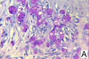
Gastric tumors in dogs are an uncommon pathology within neoplasms, and can present themselves by several factors such as the presence of antigens, in this context, this material addresses morphological and histopathological aspects of a case of gastric mucinous adenocarcinoma associated with helicobacteriosis and paracoccidioidomycosis in a 12-year-old chow-chow dog who had recurrent emesis with decreased appetite, and that she underwent partial gastrectomy. Ultrasound examination showed hyperechoic lesions in the gastric mucosa and sharply thickened walls, so endoscopy was requested, which demonstrated the existence of multiple ulcerative lesions and mucosa containing metaplasias, necrotic points and hemorrhagic foci. Next, a biopsy of the gastric mucosa was performed, in which 16 fragments were sent for histopathological analysis, and neoplastic proliferation of epithelial origin with malignant biological behavior was found. It was also possible through this analysis to diagnose the patient with mucinous adenocarcinoma, which is classified as invasive due to its disposition and tissue organization in the stomach; Chronic inflammatory reactions were also found associated with structures compatible with helicobacteriosis and paracoccidioidomycosis, possibly also related to dysplasia. This case report aims to correlate the tumor diagnosis with fungal and bacterial infection, seeking to associate the emergence and progression of the pathology with the aforementioned antigens (Helicobacter spp. and Paracoccidioides spp.), based on other literature that discusses similar cases and/or the profile of the antigens described.
References
Alberti, T. S., Venancio, F. R., Zamboni, R., Scheid, H. V, Ladeira, R. S., Sallis, E. S. V, & Schild, A. L. (2021). Canine gastric mucinous adenocarcinoma. Research, Society and Development, 10(11). https://doi.org/10,33448/rsd-v10i11.19146.
Baj, J., Korona-Glowniak, I., Forma, A., Maani, A., Sitarz, E., Rahnama-Hezavah, M., Radzikoska, E., & Portincasa, P. (2020). Mecanismos da transição rpitelial-mesenquimal e microambiente tumoral no câncer gástrico induzido por Helicobacter pylori. Células, 9(4), 1055. https://doi.org/10.3390/cells9041055.
Balakrishnan, A., & Drobatz, K. J. (2013). Management of urinary tract emergencies in small animals. Veterinary Clinics: Small Animal Practice, 43(4), 843–867. https://doi.org/10.1016/j.cvsm.2013.03.013.
Campos, M. S., Araújo, M. A., Souza, N. C., Santos, S. F., Souza, V. O., & Carneiro, R. L. (2023). Sarcoma indiferenciado por provável infiltrado de Helicobacter spp. na região antro pilórica: Relato de caso. PUBVET, 17(8), e1435. https://doi.org/10.31533/pubvet.v17n8e1435.
Choi, J. S., Kim, M. A., Lee, H. E., Lee, H. S., & Kim, W. H. (2009). Mucinous gastric carcinomas: clinicopathologic and molecular analyses. Cancer, 115(15).
Cleary, K. R. (1987). Tumors of the alimentary tract. Clinics in Laboratory Medicine, 7(1). https://doi.org/10.1016/s0272-2712(18)30762-5.
Crow, S. E. (1985). Tumors of the alimentary tract. In The Veterinary clinics of North America. Small animal practice (Vol. 15, Issue 3). https://doi.org/10.1016/S0195-5616(85)50059-5.
Czajkowski, P. S., Parry, N. M., Wood, C. A., Casale, S. A., Phipps, W. E., Mahoney, J. A., Spector, D. I., Price, L. L., & Berg, J. (2022). Outcome and Prognostic Factors in Cats Undergoing Resection of Intestinal Adenocarcinomas: 58 Cases (2008–2020). Frontiers in Veterinary Science, 9. https://doi.org/10.3389/fvets.2022.911666
Falchetti, M., Saieva, C., Lupi, R., Masala, G., Rizzolo, P., Zanna, I., Ceccarelli, K., Sera, F., Mariani-Costantini, R., Nesi, G., Palli, D., & Ottini, L. (2008). Gastric cancer with high-level microsatellite instability: target gene mutations, clinicopathologic features, and long-term survival. Human Pathology, 39(6). https://doi.org/10.1016/j.humpath.2007.10.024
Fossum, T. W. (2021). Cirurgia de pequenos animais (3ed.). Elsevier Editora.
Gabellini, G. C., Martinez, R., Ejima, F. H., Saldanha, J. C., Modena, J. L. P., Velludo, M. A. L., & Figueiredo, J. F. C. (1992). Paracoccidioidomicose do estômago: Relato de caso e considerações sobre a patogenia desta lesão. Arquivos de Gastroenterologia, 29(4), 147–152.
Galvão, J. F. B., Pressler, B. M., Freeman, L. J., Rohleder, J. J., Burgess, R. C. F., & Ramos-Vara, J. A. (2009). Mucinous gastric carcinoma with abdominal carcinomatosis and hypergastrinemia in a dog. Journal of the American Animal Hospital Association, 45(4), 197–202. https://doi.org/10.5326/0450197.
Hardas, A., Suárez-Bonnet, A., Beck, S., Becker, W. E., Ramírez, G. A., & Priestnall, S. L. (2021). Canine gastric carcinomas: A histopathological and immunohistochemical study and similarities with the human counterpart. Animals, 11(5). https://doi.org/10.3390/ani11051409
Ishaq, S., & Nunn, L. (2015). Helicobacter pylori and gastric cancer: a state-of-the-art review. Gastroenterology and Hepatology from Bed to Bench, 8(Suppl 1), 6–14.
Kleperer, P. (1949). Tumors of the alimentary tract. Hawaii Medical Journal, 8(3). https://doi.org/10.1002/9781119181200.ch13.
Ladeira, M. S. P., Salvadori, D. M. F., & Rodrigues, M. A. M. (2003). Biopatologia do Helicobacter pylori. Jornal Brasileiro de Patologia e Medicina Laboratorial, 39, 335–342. https://doi.org/10.1590/S1676-24442003000400011.
Lima, L R S, Silva, B. P., & Gaeta, F. A. (2016). Carcinoma gástrico em cão: endoscopia e histopatológico – Relato de caso. Revista de Educação Continuada em Medicina Veterinária e Zootecnia Do CRMV-SP, 14(2), 57–57.
Lourenço, T. V., & Carvalho, D. (2024). Adenocarcinoma pulmonar em felinos: Revisão. PUBVET, 18(1), e1532. https://doi.org/10.31533/pubvet.v18n01e1532.
Meuten, D. J. (2016). Tumors in domestic animals. John Wiley & Sons.
Millington, M. A., Nishioka, S. A., Martins, S. T., Santos, Z. M. G., Lima Júnior, F. E. F., & Alves, R. V. (2018). Paracoccidioidomicose: Abordagem histórica e perspectivas de implantação da vigilância e controle. Epidemiologia e Serviços de Saúde: Revista do Sistema Único de Saúde do Brasil, 27(spe). https://doi.org/10.5123/S1679-49742018000500002.
Mladenova-Hristova, I., Grekova, O., & Patel, A. (2017). Zoonotic potential of Helicobacter spp. In Journal of Microbiology, Immunology and Infection (Vol. 50, Issue 3, pp. 265–269). https://doi.org/10.1016/j.jmii.2016.11.003.
Moulton, J. E., Tscharner, C. Von, & Schneider, R. (1981). Classification of lung carcinomas in the dog and cat. Veterinary Pathology, 18(4), 513–528. https://doi.org/10.1177/030098588101800409.
Okubo, B. M., Ricci-Azevedo, R., Zobiole, N. N., Buccini, D. F., & Moreno, S. E. (2017). Prevalência de Helicobacter spp. em cães de Campo Grande-MS. Ciência Animal Brasileira, 18, 1–10. https://doi.org/10.1590/1089-6891v18e-17286.
Owen, L. N. (1980). TNM Classification of tumours in domestic animals. World Health Organization.
Paoloni, M. C., Penninck, D. G., & Moore, A. S. (2002). Ultrasonographic and clinicopathologic findings in 21 dogs with intestinal adenocarcinoma. Veterinary Radiology & Ultrasound, 43(6), 562–567. https://doi.org/10.1111/j.1740-8261.2002.tb01050.x.
Saito, T., Nibe, K., Chambers, J. K., Uneyama, M., Nakashima, K., Ohno, K., Tsujimoto, H., Uchida, K., & Nakayama, H. (2020). A histopathological study on spontaneous gastrointestinal epithelial tumors in dogs. Journal of Toxicologic Pathology, 33(2). https://doi.org/10.1293/tox.2019-0076
Sepulvida, M. B. C., Stelini, R. F., Pompeu. T R C, & Teixeira, M. A. B. T. (2013). Gastric ulcer perforation credited to paracoccidioidomycosis. Case report with autopsy. Revista Da Sociedade Brasileira e Clínicas Médicas, 11(4), 360–363.
Vasconcellos, M. B., Favaris, J. W. de S., Campos, M. M., Chavaglia, L. C. R., Ribeiro, T. B. B., Souza, M. L. de, & Cerruti, C. H. (2022). Adenocarcinoma mucinoso de apêndice: estudo de caso com abordagem intraoperatória. Revista Eletrônica Acervo Saúde, 15(6). https://doi.org/10.25248/reas.e9911.2022
Wagner, F., Oro, R., Campos, V. Z., Rocha, B. L., Dalegrave, S., Wilmsen, M. O., & Mazzuco, J. T. (2021). Leiomiossarcoma gástrico canino: Relato de caso. PUBVET, 15(11), 1–6. https://doi.org/10.31533/pubvet.v15n11a964.1-6.
Zachary, J. F., McGavin, D., & McGavin, M. D. (2012). Bases da patologia em veterinária. Elsevier Brasil.
Zortéa, M. F. M., Lemos, V. Z., Bins, I. O. G., Sede, S. C., Fonseca, I. F., Orzil, I. de P., Rossoni Júnior, J. V., & Pereira, C. M. (2024). Adenoma de glândula perirenal - Relato de caso. Revista Foco, 17(3), e3560. https://doi.org/10.54751/revistafoco.v17n3-006.
Downloads
Published
Issue
Section
License
Copyright (c) 2025 Dra. Carla Sordi Furlanetto, Dr. Luiz Henrique Grisa, Dr. Wander Gawenda Ziger, Julia Da R. Tavares, Roberto Daniel Pizzolatto, Dra. Leticia Maria Santos Silva

This work is licensed under a Creative Commons Attribution 4.0 International License.
Você tem o direito de:
Compartilhar — copiar e redistribuir o material em qualquer suporte ou formato
Adaptar — remixar, transformar, e criar a partir do material para qualquer fim, mesmo que comercial.
O licenciante não pode revogar estes direitos desde que você respeite os termos da licença. De acordo com os termos seguintes:
Atribuição
— Você deve dar o crédito apropriado, prover um link para a licença e indicar se mudanças foram feitas. Você deve fazê-lo em qualquer circunstância razoável, mas de nenhuma maneira que sugira que o licenciante apoia você ou o seu uso. Sem restrições adicionais
— Você não pode aplicar termos jurídicos ou medidas de caráter tecnológico que restrinjam legalmente outros de fazerem algo que a licença permita.


