
Epibulbar melanocytoma in a domestic cat
Case report
DOI:
https://doi.org/10.31533/pubvet.v19n02e1729Keywords:
Enucleation, melanocytic lesions, ocular neoplasms, veterinary pathologyAbstract
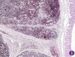
Melanocytic lesions are the most common cause of ocular neoplasia observed in cats. Melanocytomas are melanocytic lesions with benign behavior. What differentiates lesions with high malignant potential from those with low malignant potential is the histopathological analysis of the material, since it is not possible to define their potential based on the macroscopic view of the lesion. The present study aimed to report a case of epibulbar melanocytoma in an adult, spayed, mixed-breed cat, with a blackish-colored increase in volume in the epibulbar region of the upper eyelid region of the left eye. A surgical approach regarding the tumor was chosen, with transconjunctival enucleation of the affected eye, which underwent histopathological analysis, and the diagnosis of melanocytoma was confirmed. Given the apparent similarity between malignant and benign ocular melanocytic lesions, it is concluded that the analysis of enucleated material is necessary when an ocular melanocytic lesion is suspected, since the diagnosis can define the postoperative conduct and prognosis.
References
August, J. R. (2011). Medicina interna de felinos. Elsevier.
Bojrab, M. J. (2014). Mecanismos das doenças em cirurgia de pequenos animais (Vol. 1). Roca.
Daleck, C. R., Fonseca, C. S., & Canola, J. C. (2016). Oncologia em cães e gatos. Roca.
Kanai, K., Kanemaki, N., Matsuo, S., Ichikawa, Y., Okujima, H., & Wada, Y. (2006). Excision of a feline limbal melanoma and use of nictitans cartilage to repair the resulting corneoscleral defect. Veterinary Ophthalmology, 9(4). https://doi.org/10.1111/j.1463-5224.2006.00452.x.
Kayes, D., & Blacklock, B. (2022). Feline uveal melanoma review: Our current understanding and recent research advances. In Veterinary Sciences, 9(2): 9–46. https://doi.org/10.3390/vetsci9020046.
Schobert, C. S., Labelle, P., & Dubielzig, R. R. (2010). Feline conjunctival melanoma: Histopathological characteristics and clinical outcomes. Veterinary Ophthalmology, 13(1), 43–46. https://doi.org/10.1111/j.1463-5224.2009.00758.x.
Silva, K. L. F. (2013). Estudo da proliferação celular em tumores melanocíticos caninos. In Departamento de Veterinária: Vol. Master of Science.
Turner, S. (2010). Oftalmología de pequeños animales. Elselvier Saunders.
Vail, D. M., Thamm, D. H., & Liptak, J. M. (2019). Withrow and MacEwen’s Small Animal Clinical Oncology-E-Book. Elsevier Health Sciences.
Wang, A. L., & Kern, T. (2015). Melanocytic ophthalmic neoplasms of the domestic veterinary species: A review. In Topics in Companion Animal Medicine, 30(4):148–157. https://doi.org/10.1053/j.tcam.2015.06.001.
Withrow, S. J., Page, R., & Vail, D. M. (2020). Small animal clinical oncology. Elsevier Health Sciences. https://doi.org/10.1201/9781315381855.
Downloads
Published
Issue
Section
License
Copyright (c) 2025 Tatiana de França Sales, Fernanda Conde, Franceliusa Delys de Oliveira, Camila Sabaudo Alves

This work is licensed under a Creative Commons Attribution 4.0 International License.
Você tem o direito de:
Compartilhar — copiar e redistribuir o material em qualquer suporte ou formato
Adaptar — remixar, transformar, e criar a partir do material para qualquer fim, mesmo que comercial.
O licenciante não pode revogar estes direitos desde que você respeite os termos da licença. De acordo com os termos seguintes:
Atribuição
— Você deve dar o crédito apropriado, prover um link para a licença e indicar se mudanças foram feitas. Você deve fazê-lo em qualquer circunstância razoável, mas de nenhuma maneira que sugira que o licenciante apoia você ou o seu uso. Sem restrições adicionais
— Você não pode aplicar termos jurídicos ou medidas de caráter tecnológico que restrinjam legalmente outros de fazerem algo que a licença permita.


