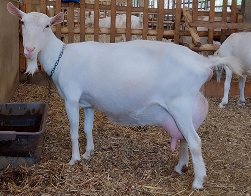
Junctional rhythm during trans-anesthetic monitoring in a goat (Capra aegagrus) with ulcerated neoformation
Case report
DOI:
https://doi.org/10.31533/pubvet.v18n12e1697Keywords:
balanced anesthesia, electrocardiogram, ruminants, farm animalsAbstract
Supraventricular ectopic beats and their rhythms can occur both in the presence and absence of heart disease, secondary to systemic diseases. Its arrhythmogenic mechanisms include enhanced automaticity, triggered activity, and anatomical changes or reentry mechanisms. The junctional region is the normal conduction pathway between atria and ventricles, and can be divided into three segments: atrioventricular node (AVN), Hissian node (HN) and bundle of His. A goat with a neoformation in the vulvar region went through an anesthetic procedure for incisional biopsy, urethral clearance and probing. As pre-anesthetic medication, midazolam (0.2 mg/kg) and morphine (0.1 mg/kg) were administered intravenously, induction with propofol (3 mg/kg), orotracheal intubation and maintenance with isoflurane. Epidural anesthesia was performed in the lumbosacral region with lidocaine (4 mg/kg) and morphine (0.1 mg/kg). Trans-anesthetic monitoring was performed with electrocardiogram (evaluating rhythm and heart rate), pulse oximetry, capnography and invasive mean arterial pressure (MAP), in addition to observing protective reflexes to monitor the anesthetic plane. Upon notice of hypotension and junctional rhythm on the electrocardiogram, 0.04 mg/kg of atropine was administered intravenously (IV), resuming sinus rhythm after application. During the change, no P wave was observed and, after the administration of atropine, the P wave appeared for a few minutes before returning to the junctional rhythm, which was maintained until the end of anesthesia and in the immediate postoperative period.
References
Barbosa, R. R., Fontenele-Neto, J. D., & Soto-Blanco, B. (2008). Toxicity in goats caused by oleander (Nerium oleander). Research in Veterinary Science, 85(2), 279–281. https://doi.org/10.1016/j.rvsc.2007.10.004.
Barretto, F. L., Ferreira, F. S., Freitas, M. V, Santos, V. S., Correa, E. S., & Carvalho, C. B. (2013). Eletrocardiografia contínua (Holter) em cães saudáveis submetidos a diferentes exercícios físicos. Arquivo Brasileiro de Medicina Veterinária e Zootecnia, 65, 1625–1634.
Basso, P. C., Raiser, A. G., Carregaro, A. B., & Muller, D. C. M. (2008). Analgesia transoperatória em cães e gatos. Clínica Veterinária, 77, 62–68.
Bastos, E. J. D., Humberto, E. C., Magalhães, A. O. C., Kamimura, R., Cunha, G. N., & Santos, M. C. (2007). Alterações histopatológicas do nó sinoatrial em cães com dilatação cardíaca. Veterinária Notícias, 13(2), 15–21.
Bastos, J. E. D., Silva, N. M., Briceño, M. P. P., Wilson, T. M., & Ronchi, A. A. M. (2020). Anatomopathological changes in canine distemper seropositive dogs and virus detection in sinoatrial nodes. Bioscience Journal, 487–495.
Brito, F. S. (2023). Eletrocardiografia ambulatorial: Sistema Holer e monitor de eventos (Looper) - Interpretação de análises. In Procardiol: Programa de atualização em cardiologia. https://doi.org/10.5935/978-85-514-1162-9.c0002
Carvalho, C. F., Tudury, E. A., Neves, I. V, Fernandes, T. H. T., Gonçalves, L. P., & Salvador, R. (2009). Eletrocardiografia pré-operatória em 474 cães. Arquivo Brasileiro de Medicina Veterinária e Zootecnia, 61(3), 590–597. https://doi.org/10.1590/s0102-09352009000300011.
Cassiano Neto, J. (1976). Fisiopatolia da disfunção isiopatologia da disfunção do nódulo sinoatrila. Arquivos Brasileiros de Cardiologia, 29(2).
Cunningham, J. (2011). Tratado de fisiologia veterinária. Guanabara Koogan.
Fantoni, D. T., & Cortopassi, S. R. G. (2009). Anestesia em cães e gatos. Roca.
Fantoni, D. T., & Mastrocinque, S. (2005). Analgesia preventiva. In P. E. Otero (Ed.), Dor: Avaliação e tratamento em pequenos animais (pp. 76–80). Interbook.
French, A. (2004). Electrocardiography in the dog and cat. In Irish Veterinary Journal (Vol. 57, Issue 7).
Hamlin, R. L., Glower, D. D., & Pimmel, R. L. (1984). Genesis of QRS in the ruminant: Graphic simulation. American Journal of Veterinary Research, 45(5), 938–941.
Holmes, J. R. (1990). Electrocardiography in the diagnosis of common cardiac arrhythmias in the horse. Equine Veterinary Education, 2(1), 24–27. https://doi.org/10.1111/j.2042-3292.1990.tb01373.x.
Klein, B. G. (2014). Cunningham Tratado de Fisiologia Veterinária. Elsevier.
Luna, S. P. L., & Carregaro, A. B. (2019). Anestesia e analgesia em equídeos, ruminantes e suínos. Editora MedVet. https://doi.org/10.36440/recmvz.v1i1.3392.
Muir, W. W., & Hubbell, J. A. E. (2001). Manual de anestesia veterinária. Artmed Editora.
Mukherjee, J., Mohapatra, S. S., Jana, S., Das, P. K., Ghosh, P. R., Das, K., & Banerjee, D. (2020). A study on the electrocardiography in dogs: Reference values and their comparison among breeds, sex, and age groups. Veterinary World, 13(10), 2216. https://doi.org/10.14202/vetworld.2020.2216-2220.
Oliveira, G. A. C. (2012). Continuous electrocardiography in dogs and cats. In A Bird’s-Eye View of Veterinary Medicine. https://doi.org/10.5772/33636.
Pogliani, F. C., Birgel, E. H., Monteiro, B. M., Grisi Filho, J. H. H., & Raimondo, R. F. S. (2013). The normal electrocardiogram in the clinically healthy Saanen goats. Pesquisa Veterinaria Brasileira, 33(12), 1478–1482. https://doi.org/10.1590/S0100-736X2013001200014.
Reddy, B. S., Venkatasivakumar, R., Sivajothi, S., & Reddy, Y. V. P. (2013). Electrocardiographic abnormalities in young healthy sheep and goats. International Journal of Biological Research, 2(1). https://doi.org/10.14419/ijbr.v2i1.2252.
Skarda, R. T., Muir, W. W., & Hubbell, J. A. E. (2009). Local anesthetic drugs and techniques. Equine Anesthesia, 1(1), 210–242. https://doi.org/10.1111/eve.13235.
Szabuniewicz, M., & Clark, D. R. (1967). Analysis of the electrocardiograms of 100 normal goats. American Journal of Veterinary Research, 28(123), 511–516.
Tilley, L. P. (1985). Essentials of canine and feline electrocardiography: interpretation and treatment. Lea & Febiger.
Willis, R., Oliveira, P., & Mavropoulou, A. (2018). Guide to canine and feline electrocardiography. In Guide to Canine and Feline Electrocardiography. https://doi.org/10.1002/9781119254355.

Downloads
Published
Issue
Section
License
Copyright (c) 2024 Júlia Ferraz Cereda Martinez, Juliana Rizerio Moncayo, Maria Fernanda Almeida Cardoso, Beatriz Sanches Rosa, Zahi Eni dos Santos Souza, Andressa de Fátima kotleski Thomaz de Lima

This work is licensed under a Creative Commons Attribution 4.0 International License.
Você tem o direito de:
Compartilhar — copiar e redistribuir o material em qualquer suporte ou formato
Adaptar — remixar, transformar, e criar a partir do material para qualquer fim, mesmo que comercial.
O licenciante não pode revogar estes direitos desde que você respeite os termos da licença. De acordo com os termos seguintes:
Atribuição
— Você deve dar o crédito apropriado, prover um link para a licença e indicar se mudanças foram feitas. Você deve fazê-lo em qualquer circunstância razoável, mas de nenhuma maneira que sugira que o licenciante apoia você ou o seu uso. Sem restrições adicionais
— Você não pode aplicar termos jurídicos ou medidas de caráter tecnológico que restrinjam legalmente outros de fazerem algo que a licença permita.


