
Urolithiasis in a Pug
Case report
DOI:
https://doi.org/10.31533/pubvet.v18n10e1667Keywords:
Cystotomy, dog, urolithiasis, struvite urolithsAbstract
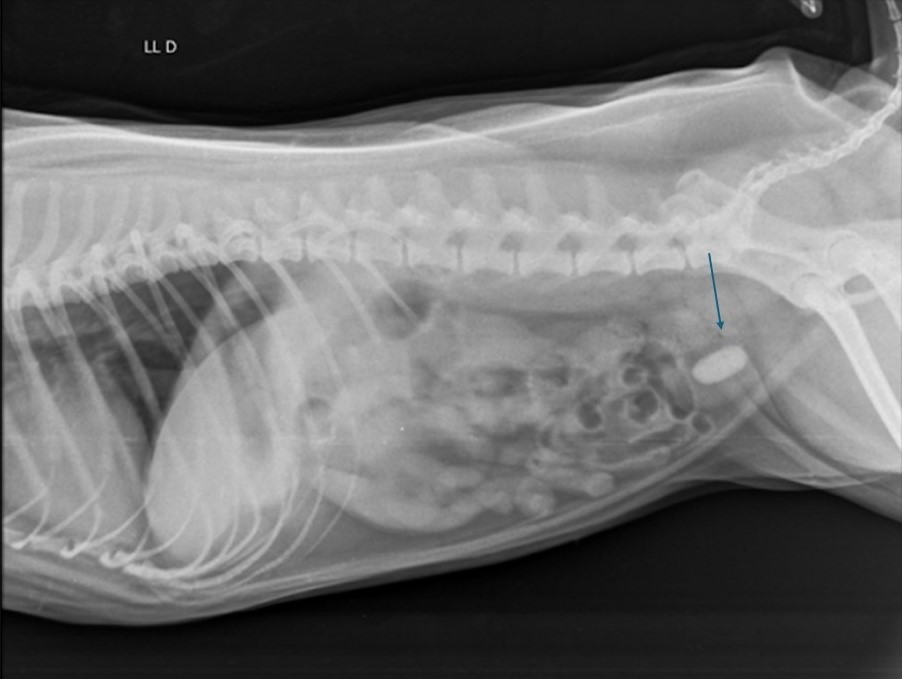
The objective of this paper was to present a case of urolithiasis in a two-year old Pug weighing ten kilos, with a history of food allergy/atopic dermatitis. Upon physical examination, the animal presented mild abdominal pain upon palpation. The tutor indicated emesis 2 days ago. Upon ultrasound examination, it was possible to identify the presence of a 2.2 cm urolith in the bladder, in addition to cystitis in the urinary tract. The animal underwent surgical treatment, with cystotomy being the most suitable for this type of case. From the case report, it was found that racial or even familial predisposition was the main cause of urolith formation. In addition, the chances of urolithiasis occurring are greater in small breed dogs.
References
Caporali, E. H. G., Phillips, H., Underwood, L., & Selmic, L. E. (2015). Risk factors for urolithiasis in dogs with congenital extrahepatic portosystemic shunts: 95 cases (1999–2013). Journal of the American Veterinary Medical Association, 246(5), 530–536. https://doi.org/10.2460/javma.246.5.530.
Ferraz, M. L., Eurides, A. C., Machado, B. R., Baioco, L. H. S., Rossi, A. D., & Wiecheteck, V. S. (2020). Urolitíase em um cão da raça Pug. PUBVET, 14(9), 1–5. https://doi.org/10.31533/pubvet.v14n9a641.1-5
Fossum, T. W. (2021). Cirurgia de pequenos animais (3ed.). Elsevier Editora.
Goloni, C., Bonder, B. S. A., Senhorello, I. L. S., Tinucci-Costa, M., & Carciofi, A. C. (2018). Dissolução de urólito de estruvita por meio de manejo nutricional e antibioticoterapia em cão: Relato de caso. Ars Veterinaria, 34(3), 135–140. https://doi.org/10.15361/2175-0106.2018v34n3p135-140.
Houston, D. M., & Moore, A. E. P. (2009). Canine and feline urolithiasis: examination of over 50 000 urolith submissions to the Canadian veterinary urolith centre from 1998 to 2008. The Canadian Veterinary Journal, 50(12), 1263–1268.
Inkelmann, M. A., Kommers, G. D., Trost, M. E., Barros, C. S. L., Fighera, R. A., Irigoyen, L. F., & Silveira, I. P. (2012). Urolitíase em 76 cães. Pesquisa Veterinária Brasileira, 32(3), 247–253. https://doi.org/10.1590/S0100-736X2012000300012.
Koehler, L. A., Osborne, C. A., Buettner, M. T., Lulich, J. P., & Behnke, R. (2009). Canine uroliths: frequently asked questions and their answers. Veterinary Clinics of North America: Small Animal Practice, 39(1), 161–181.
Kopecny, L., Palm, C. A., Segev, G., & Westropp, J. L. (2021). Urolithiasis in dogs: Evaluation of trends in urolith composition and risk factors (2006‐2018). Journal of Veterinary Internal Medicine, 35(3), 1406–1415. https://doi.org/10.1111/jvim.16114.
Kuntz, J. A., Berent, A. C., Weisse, C. W., & Bagley, D. H. (2015). Double pigtail ureteral stenting and renal pelvic lavage for renal-sparing treatment of obstructive pyonephrosis in dogs: 13 cases (2008–2012). Journal of the American Veterinary Medical Association, 246(2). https://doi.org/10.2460/javma.246.2.216.
Lazzarotto, J. J. (2000). Doença do trato urinário inferior dos felinos associada aos cristais de estruvita. Revista Da Faculdade de Zootecnia, Veterinária e Agronomia, 7–8(1), 58–64.
Leite, A. C., Almeida, A. C., Araújo, A. H. B., Schultz, E. B., Araújo, B. P. G., Araújo, S. V. S. de C., & Reis, R. C. S. (2020). Dieta natural no tratamento de cão acometido com recorrentes urólitos de oxalato de cálcio: Relato de caso. PUBVET, 14(11), 1–4. https://doi.org/10.31533/pubvet.v14n11a681.1-4.
Lulich, J. P., Osborne, C. A., & Albasan, H. (2010). Canine and feline urolithiasis: diagnosis, treatment, and prevention. In Nephrology and Urology of Small Animals (pp. 687–706). Wiley. https://doi.org/10.1002/9781118785546.ch69.
Marcodes, I., Rodrigues, N., Massoni, E. A., Merida, F., & Domingues, L. M. (2022). Urolitíase em suíno: Relato de caso. PUBVET, 16(11), 1–6. https://doi.org/10.31533/pubvet.v16n11a1273.1-6.
Martins, A. G. C., & Oliveira, M. K. R. (2021). Urolitíase em cães: Relato de caso.
Mendoza-López, C. I., Angel-Caraza, J., Aké-Chiñas, M. A., Quijano-Hernández, I. A., & Barbosa-Mireles, M. A. (2020). Canine silica urolithiasis in Mexico (2005–2018). Veterinary Medicine International, 2020. https://doi.org/10.1155/2020/8883487.
Monferdini, R. P., & Oliveira, J. (2009). Manejo nutricional para cães e gatos com urolitíase–Revisão bibliográfica. Acta Veterinaria Brasilica, 3(1), 1–4.
Okafor, C. C., Lefebvre, S. L., Pearl, D. L., Yang, M., Wang, M., Blois, S. L., Lund, E. M., & Dewey, C. E. (2014). Risk factors associated with calcium oxalate urolithiasis in dogs evaluated at general care veterinary hospitals in the United States. Preventive Veterinary Medicine, 115(3–4), 217–228.
Oliveira, J. V., Almeida, M. D. S., Cavalcante, L. C., Peixoto, T. K. F., Almeidas, B. V., & Leite, A. K. R. M. (2018). Alterações clínicas e laboratoriais em uma cadela com urolitíase: Relato de caso. Revista Científica de Medicina e Veterinária, 30.
Osborne, C. A., Lulich, J. P., Kruger, J. M., Ulrich, L. K., & Koehler, L. A. (2009). Analysis of 451,891 canine uroliths, feline uroliths, and feline urethral plugs from 1981 to 2007: Perspectives from the Minnesota Urolith Center. In Veterinary Clinics of North America - Small Animal Practice (Vol. 39, Issue 1). https://doi.org/10.1016/j.cvsm.2008.09.011.
Osborne, C. A., Polzin, D. J., Kruger, J. M., Lulich, J. P., Johnston, G. R., & O’Brien, T. D. (1989). Relationship of nutritional factors to the cause, dissolution, and prevention of feline uroliths and urethral plugs. In The Veterinary clinics of North America. Small animal practice (Vol. 19, Issue 3, pp. 561–581). https://doi.org/10.1016/S0195-5616(89)50061-5.
Osborne, C. A., Polzin, D. J., Lulich, J. P., Kruger, J. M., Johnston, G. R., O’Brien, T. D., & Felice, L. J. (1989). Relationship of nutritional factors to the cause, dissolution, and prevention of canine uroliths. In The Veterinary clinics of North America. Small animal practice (Vol. 19, Issue 3, pp. 583–619). https://doi.org/10.1016/S0195-5616(89)50062-7.
Osborne, D., Carl, A., Lulich, D., & Jody P. (2008). Changing paradigms in diagnosis and treatment of urolithiasis. Veterinary Clinical Small Animal, 39.
Oyafuso, M. K. (2008). Estudo retrospectivo e prospectivo da urolitíase em cães. Universidade de São Paulo.
Oyafuso, M. K., Kogika, M. M., Waki, M. F., Prosser, C. S., Cavalcante, C. Z., & Wirthl, V. A. B. F. (2010). Urolitíase em cães: Avaliação quantitativa da composição mineral de 156 urólitos. Ciência Rural, 40(1), 102–108. https://doi.org/10.1590/s0103-84782010000100017.
Picavet, P., Detilleux, J., Verschuren, S., Sparkes, A., Lulich, J., Osborne, C., Istasse, L., & Diez, M. (2007). Analysis of 4495 canine and feline uroliths in the Benelux. A retrospective study: 1994-2004. Journal of Animal Physiology and Animal Nutrition, 91(5–6). https://doi.org/10.1111/j.1439-0396.2007.00699.x.
Raditic, D. M. (2015). Complementary and integrative therapies for lower urinary tract diseases. Veterinary Clinics: Small Animal Practice, 45(4), 857–878. https://doi.org/10.1016/j.cvsm.2015.02.009.
Rick, G. W., Conrad, M. L. H., Vargas, R. M., Machado, R. Z., Lang, P. C., Serafini, G. M. C., & Bones, V. C. (2017). Urolitíase em cães e gatos. PUBVET, 11, 646–743.
Rodríguez Díaz, M. (2016). Aportaciones al conocimiento de la urolitiasis canina y felina en España. In Repositorio Institucional.
Roe, K., Pratt, A., Lulich, J., Osborne, C., & Syme, H. M. (2012). Analysis of 14,008 uroliths from dogs in the UK over a 10‐year period. Journal of Small Animal Practice, 53(11), 634–640. https://doi.org/10.1111/j.1748-5827.2012.01275.x.
Silva Filho, E., Prado, T., Ribeiro, R., & Fortes, R. (2013). Urolitíase canina. Enciclopédia Biosfera, 9(17), 2517–2536.
Stevenson, A., & Rutgers, C. (2006). Nutritional management of canine urolithiasis. In P. Pibot, D. Biouge, & V. Elliot (Eds.), Encyclopedia of canine clinical nutrition (pp. 284–315). Royal Canin Missouri.
Tavares, T. C., Teixeira, N. D., & Oliveira Júnior, I. M. (2023). Urolitíase em cão da raça Pug: Relato de caso. PUBVET, 17(2), e1341. https://doi.org/10.31533/pubvet.v17n02a1341.
Downloads
Published
Issue
Section
License
Copyright (c) 2024 Raquel da Mata Mazzonetto Pinto

This work is licensed under a Creative Commons Attribution 4.0 International License.
Você tem o direito de:
Compartilhar — copiar e redistribuir o material em qualquer suporte ou formato
Adaptar — remixar, transformar, e criar a partir do material para qualquer fim, mesmo que comercial.
O licenciante não pode revogar estes direitos desde que você respeite os termos da licença. De acordo com os termos seguintes:
Atribuição
— Você deve dar o crédito apropriado, prover um link para a licença e indicar se mudanças foram feitas. Você deve fazê-lo em qualquer circunstância razoável, mas de nenhuma maneira que sugira que o licenciante apoia você ou o seu uso. Sem restrições adicionais
— Você não pode aplicar termos jurídicos ou medidas de caráter tecnológico que restrinjam legalmente outros de fazerem algo que a licença permita.


