
Determination of tracheal diameter reference values compared to the thoracic entry in clinically normal Shih-Tzu dogs
DOI:
https://doi.org/10.31533/pubvet.v18n10e1662Keywords:
Particularities, radiography, Shih-tzuAbstract
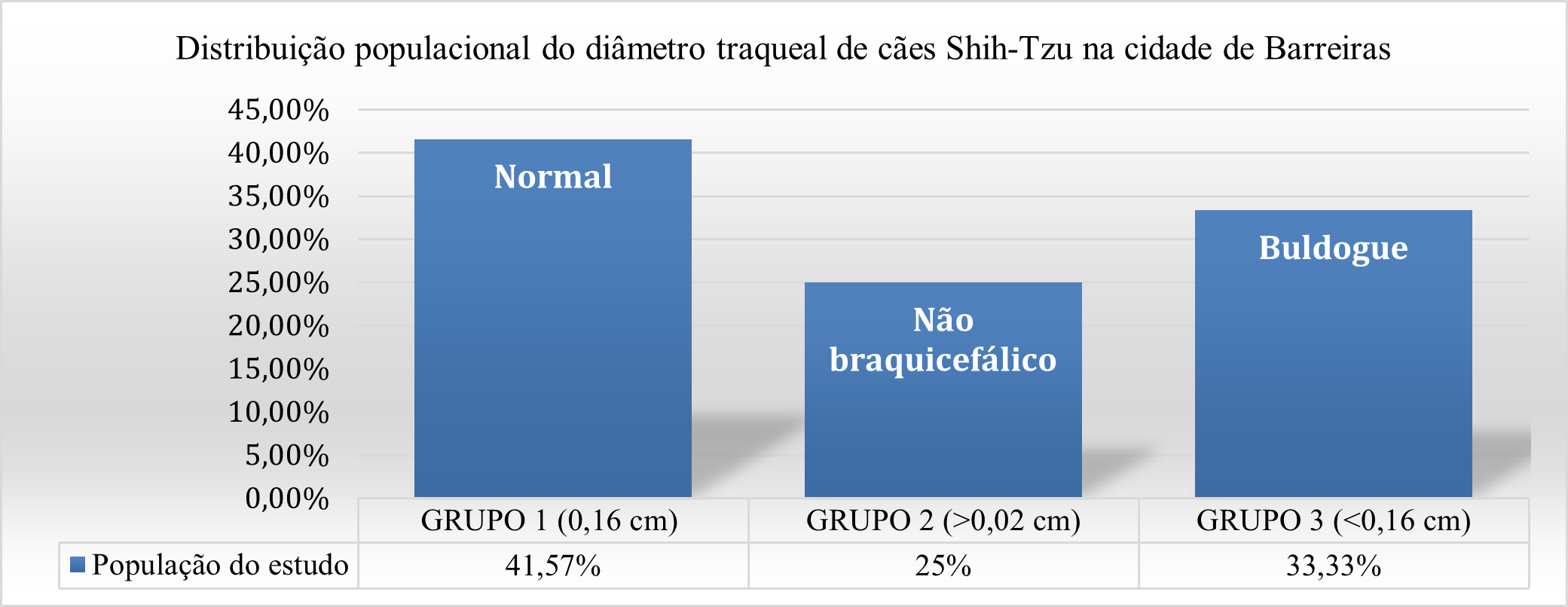
The trachea has the important function of connecting the upper and lower respiratory systems, it has a flexible tubular structure, containing cartilaginous rings along its entire length. Imaging technology makes it possible to study this organ, and can then diagnose dysfunctions and natural thinning of its diameter, as occurs with the Shih-tzu breed (brachycephalic). The study with brachycephalic breeds aims to define racial characteristics in relation to the aforementioned organ through radiographic examination. Image analysis was performed with 24 Shih-tzus of both sexes, 1 to 9 years old. In the study, 41.67% of the dogs of both sexes obtained values within the references studied; 25% of the animals obtained values equal and above 0.2 cm, fitting the parameters for non-brachycephalic dogs, and 33.33% were below 0.16, fitting the bulldog breed. Further studies are needed for a better clinical understanding of the particularities of this breed.
References
Bezerra, H. P., & Marinho, R. S. L. (2018). Alterações anatômicas primárias das vias respiratórias em cães braquicefálicos: Revisão de literatura.
Canola, J. C., Medeiros, F. P., & Canola, P. A. (2016). Radiografia convencional, ultrassonografia, tomografia e ressonância magnética. In C. R. Daleck, A. B. De Narde, & S. Rodaski (Eds.), Oncologia em cães e gatos (pp. 133–135). Roca, Brasil.
Cavinatto, C. C., Armando, A. P. R. N., Cruz, L. K. S., Lima, E. M. M., & Santana, M. I. S. (2016). Descrição anatômica de esqueletos de papagaios do gênero Amazona através da utilização de radiografias. Pesquisa Veterinária Brasileira, 36(2), 123–130.
Coyne, B. E., & Fingland, R. B. (1992). Hypoplasia of the trachea in dogs: 103 cases (1974-1990). Journal of the American Veterinary Medical Association, 201(5), 768–772. https://doi.org/10.2460/javma.1992.201.05.768.
Dörner, N. L., Santos, A. P. L., Silva, L. K. L., & Martins, M. A. S. (2021). Uso da seleção como técnica de melhoramento genético aplicada em cães: Estudo nos canis dos municípios de São Luis e São José de Ribamar - MA. In Geração e difusão de conhecimento científico na zootecnia 2. https://doi.org/10.22533/at.ed.2912123116.
Harvey, C. E., & Fink, E. A. (1982). Tracheal diameter: Analysis of radiographic measurements in brachycephalic and nonbrachycephalic dogs. Journal of the American Animal Hospital Association, 18(4), 570–576.
Kealy, J., Graham, J., & McAllister, H. (2016). Radiografia e ultrassonografia do cão e do gato. Elsevier.
Mendes, H. L. C., Costa, G. P., Rocha, D. O. A. C., Silva, D. P. C., & Souza, J. H. B. (2022). Colapso de traqueia em um canino da raça Spitz Alemão (I Congresso Nacional de Especialidades Veterinárias, Ed.). https://doi.org/10.51161/convesp/6502.
Packer, R. M. A., & Tivers, M. (2015). Strategies for the management and prevention of conformation-related respiratory disorders in brachycephalic dogs. Veterinary Medicine: Research and Reports. https://doi.org/10.2147/vmrr.s60475.
Pavelski, M., Silva, D. M., & Froes, T. R. (2016). Radiografia das cavidades craniana e nasal em afecções neoplásicas em cães: características e limitações. Veterinária e Zootecnia, 23(2), 164–173.
Siedenburg, J. S., & Dupré, G. (2021). Tongue and upper airway dimensions: A comparative study between three popular brachycephalic breeds. Animals, 11(3), 1–13. https://doi.org/10.3390/ani11030662.
Souto, C. K., Martín, C. M., Ferrante, B., & Pinto, A. C. B. C. F. (2015). Métodos de diagnóstico por imagem para avaliação traqueal em pequenos animais. Revista Acadêmica Ciência Animal, 13, 111–123. https://doi.org/10.7213/academica.13.fc.ao12.
Yang, C., Trad, H. S., Mendonça, S. M., & Trad, C. S. (2013). Anomalias congênitas da veia cava inferior: revisão dos achados na tomografia computadorizada multidetectores e ressonância magnética. Radiologia Brasileira, 46(4), 227–233.
Downloads
Published
Issue
Section
License
Copyright (c) 2024 Lorrania Alcantara Ruas, Jackson Farias, Mariana Santos Campos, Tiago Oliveira Brandão, Naiane Darklei dos Santos Silva, Rodrigo Lima Carneiro

This work is licensed under a Creative Commons Attribution 4.0 International License.
Você tem o direito de:
Compartilhar — copiar e redistribuir o material em qualquer suporte ou formato
Adaptar — remixar, transformar, e criar a partir do material para qualquer fim, mesmo que comercial.
O licenciante não pode revogar estes direitos desde que você respeite os termos da licença. De acordo com os termos seguintes:
Atribuição
— Você deve dar o crédito apropriado, prover um link para a licença e indicar se mudanças foram feitas. Você deve fazê-lo em qualquer circunstância razoável, mas de nenhuma maneira que sugira que o licenciante apoia você ou o seu uso. Sem restrições adicionais
— Você não pode aplicar termos jurídicos ou medidas de caráter tecnológico que restrinjam legalmente outros de fazerem algo que a licença permita.


