Complete atrioventricular block in a dog with myxomatous mitral valve disease and echocardiographic limitations
Case report
DOI:
https://doi.org/10.31533/pubvet.v20n6e1973Keywords:
Cardiac arrhythmia, congestive heart failure, dog, echocardiographyAbstract
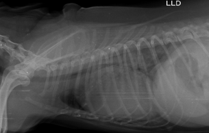
The objective of this study was to report a complex case of multimodal management and diagnostic discordance among imaging modalities in a canine patient. A 13-year-old female Poodle was presented with chronic dry cough, dyspnea, tachypnea, a grade IV/VI holosystolic murmur over the mitral valve area, and severe bradycardia. Electrocardiography revealed a supra-Hisian complete atrioventricular block (cAVB), with a ventricular rate of 49 beats per minute. Thoracic radiography demonstrated severe cardiomegaly, with a Vertebral Heart Score (VHS) of 12.4 vertebrae, a caudodorsal alveolar pulmonary pattern consistent with cardiogenic pulmonary edema, and hepatomegaly. Biochemical analysis and abdominal ultrasonography confirmed secondary congestive hepatopathy, accompanied by marked elevations in liver enzyme activities. Conversely, echocardiography revealed morphologic parameters within normal limits, including a left atrium-to-aorta (LA/Ao) ratio of 1.23 and a normalized left ventricular internal diameter in diastole (LVIDdn) of 1.87, falsely suggesting ACVIM stage B1 myxomatous mitral valve disease (MMVD), despite exhibiting hyperdynamic ejection fraction (85%) and fractional shortening (53%). Treatment for congestive heart failure (CHF) was instituted according to the American College of Veterinary Internal Medicine (ACVIM) guidelines using torasemide (0.2 mg/kg), pimobendan (0.25 mg/kg), spironolactone (2 mg/kg), and benazepril (0.5 mg/kg) every 12 hours, combined with hepatoprotective therapy. After seven days of treatment, the patient showed complete resolution of the cough and normalization of the resting respiratory pattern. In conclusion, severe bradycardia resulting from cAVB alters diastolic and capillary pressure dynamics, potentially masking conventional echocardiographic indices of cardiac remodeling in MMVD. These findings highlight the primacy of clinical assessment and the need for an integrated, multimodal diagnostic approach in dogs with complex cardiac disease.
References
Bonagura, J. D., & Schober, K. E. (2009). Can ventricular function be assessed by echocardiography in chronic canine mitral valve disease? Journal of Small Animal Practice, 50(SUPPL. 1), 12–24. https://doi.org/10.1111/j.1748-5827.2009.00803.x.
Boon, J. A. (2016). Two-Dimensional and M-Mode Echocardiography for the Small Animal Practitioner: Second Edition. In Two-Dimensional and M-Mode Echocardiography for the Small Animal Practitioner: Second Edition (2nd ed.). John Wiley & Sons Inc. https://doi.org/10.1002/9781119028574,
Boswood, A., Häggström, J., Gordon, S. G., Wess, G., Stepien, R. L., Oyama, M. A., Keene, B. W., Bonagura, J., MacDonald, K. A., & Patteson, M. (2016). Effect of pimobendan in dogs with preclinical myxomatous mitral valve disease and cardiomegaly: the EPIC study—a randomized clinical trial. Journal of Veterinary Internal Medicine, 30(6), 1765–1779. https://doi.org/10.1016/j.asams.2017.03.008.
Brito, K. G. J., Cartaxo, M. N., Silva, M. D., Pinto, L. R., Conceição, C. F. D., Chagas, C. Q. O., Mota, J. A., Dias, B. L., & Carneiro, R. L. (2025). Doença mixomatosa da valva mitral em canino: Relato de caso. PUBVET, 19(9), 1–13. https://doi.org/10.31533/pubvet.v19n09e1840.
Castro, M. G., Veado, J. C. C., Silva, E. F., & Araújo, R. B. (2009). Estudo retrospectivo ecodopplercardiográfico das principais cardiopatias diagnosticadas em cães. Arquivo Brasileiro de Medicina Veterinária e Zootecnia, 61(5), 1238–1241. https://doi.org/10.1590/S0102-09352009000500032.
Chetboul, V., Pouchelon, J. L., Menard, J., Blanc, J., Desquilbet, L., Petit, A., Rougier, S., Lucats, L., & Woehrle, F. (2017). Short-Term Efficacy and Safety of Torasemide and Furosemide in 366 Dogs with Degenerative Mitral Valve Disease: The TEST Study. Journal of Veterinary Internal Medicine, 31(6), 1629–1642. https://doi.org/10.1111/jvim.14841.
Cornell, C. C., Kittleson, M. D., Torre, P. Della, Häggström, J., Lombard, C. W., Pedersen, H. D., Vollmar, A., & Wey, A. (2004). Allometric Scaling of M-Mode Cardiac Measurements in Normal Adult Dogs. Journal of Veterinary Internal Medicine, 18(3), 311–321. https://doi.org/10.1111/j.1939-1676.2004.tb02551.x.
Detofano, J., Silva, A. C. A., Almeida, L. G., Mattei, A. S., Giordani, C., Castro, L. L. D., & Guterres, K. A. (2024). Bloqueio atrioventricular de terceiro grau associado à doença valvar mixomatosa de valva mitral, aórtica e tricúspide em um cão idoso: Relato de caso. Revista Inovavet, 2(1). https://doi.org/10.18226/30855713.v2.n1.04.
Feitosa, F. L. F. (2014). Semiologia veterinária: A arte do diagnóstico. Roca Ltda.
Guglielmini, C., Berlanda, M., Fracassi, F., Poser, H., Koren, S., & Toaldo, M. B. (2019). Electrocardiographic and echocardiographic evaluation in dogs with hypothyroidism before and after levothyroxine supplementation: A prospective controlled study. Journal of Veterinary Internal Medicine, 33(5), 1935–1942.
Häggström, J., Boswood, A., O’Grady, M., Jöns, O., Smith, S., Swift, S., Borgarelli, M., Gavaghan, B., Kresken, J. G., Patteson, M., Åblad, B., Bussadori, C. M., Glaus, T., Kovačević, A., Rapp, M., Santilli, R. A., Tidholm, A., Eriksson, A., Belanger, M. C., … DiFruscia, R. (2008). Effect of pimobendan or benazepril hydrochloride on survival times in dogs with congestive heart failure caused by naturally occurring myxomatous mitral valve disease: The QUEST study. Journal of Veterinary Internal Medicine, 22(5), 1124–1135. https://doi.org/10.1111/j.1939-1676.2008.0150.x.
Keene, B. W., Atkins, C. E., Bonagura, J. D., Fox, P. R., Häggström, J., Fuentes, V. L., Oyama, M. A., Rush, J. E., Stepien, R., & Uechi, M. (2019). ACVIM consensus guidelines for the diagnosis and treatment of myxomatous mitral valve disease in dogs. Journal of Veterinary Internal Medicine, 33(3), 1127–1140. https://doi.org/10.1111/jvim.15488. Acesso em: 2 abr. 2020.
Koster, L., Vogel, J., Springer, C. M., & Hecht, S. (2023). Radiographic lung congestion scores in dogs with acute congestive heart failure caused by myxomatous mitral valve disease. Journal of Veterinary Internal Medicine, 37(6), 1983–1991. https://doi.org/10.1111/jvim.16850.
Machado, E. O. S. L., Gonçalves, B. P., Silva, J., & Santos Filho, M. (2021). Estudo retrospectivo dos achados cardiorrespiratórios e laboratoriais de cães atendidos para avaliação pré-operatória em um serviço de cardiologia e doenças respiratórias (2017-2021). Revista Brasileira de Ciência Veterinária, 28(4). https://doi.org/10.4322/rbcv.2021.053.
Maddison, J. E., Page, S. W., & Church, D. B. (2011). Farmacologia clínica de pequenos animais. In G. A. Philip & A. F. Thomas (Eds.), Medicamentos e Reprodução (pp. 520–537). Elsevier Brasil.
Margareth, H. (2017). Farmacologia Ilustrada. In Экономика Региона.
Mendonça, D. A., Paiva, J. P., Knackfuss, F., Bendas, A., & Alberigi, B. (2022). Risk of arrhythmias in dogs with structural heart disease. Pesquisa Veterinária Brasileira, 42, e07153. https://doi.org/10.1590/1678-5150-PVB-7153.
Noszczyk-Nowak, A., Michałek, M., Kałuza, E., Cepiel, A., & Pasławska, U. (2017). Prevalence of arrhythmias in dogs examined between 2008 and 2014. Journal of Veterinary Research, 61(1), 103–110. https://doi.org/10.1515/jvetres-2017-0013.
Raffa, R., Rawls, S., & Portyansky, E. (2008). Farmacología ilustrada. Netter.
Saini, N., Uppal, S. K., & Anand, A. (2023). Radiographic Assessment of Dogs with Congestive Heart Failure. Indian Journal of Animal Research, 57(7), 946–951. https://doi.org/10.18805/IJAR.B-4352.
Santilli, R., Moise, S., & Pariaut, R. (2018). Electrocardiography of the dog and cat. Diagnosis of arrhythmias. In Angewandte Chemie International Edition, 6(11), 951–952. (Vol. 1). Edra S.p.A.
Silva, A. A., Ferreira, C. G. S., Souza, C. G., & Balero, N. M. (2023). Cardiologia veterinária: Avanços em diagnóstico e tratamentos. In Cardiologia Veterinária: Avanços em Diagnóstico e Tratamento. https://doi.org/10.59283/ebk-978-65-85898-01-0.
Silva, C. E. V., & Camacho, A. A. (2005). Alterações ecocardiográficas em cães sob tratamento prolongado com doxorrubicina. Arquivo Brasileiro de Medicina Veterinária e Zootecnia, 57(3). https://doi.org/10.1590/s0102-09352005000300004.
Silva, G. X., Oliveira, B. S. M., & Magalhães, S. T. A. (2025). Ecocardiografia na avaliação cardíaca de pequenos animais: aplicações, benefícios e limitações. PUBVET, 19(6), 1–7. https://doi.org/10.31533/pubvet.v19n06e1784.
Spinosa, H. S. S., Górniak, S. L., & Bernardi, M. M. (2017). Farmacologia aplicada à medicina veterinária. Koogan Guanabara.
Thrall, D. E. (2019). Diagnóstico de radiologia veterinária. Guanabara Koogan S.A.
Toaldo, M. B., Romito, G., Cipone, M., Diana, A., & Tursi, M. (2017). Electrocardiographic, echocardiographic, and left atrial strain imaging features of a dog with atrial flutter and third-degree atrioventricular block. Journal of Veterinary Cardiology, 19(5), 462–468. https://doi.org/10.1016/j.jvc.2017.08.004.
Tripathi, K. D. (2013). Farmacologia médica. Guanabara - Koogan.
Downloads
Published
Issue
Section
License
Copyright (c) 2026 Luís Roberto Pires, José Ribamar da Silva Júnior

This work is licensed under a Creative Commons Attribution 4.0 International License.
Você tem o direito de:
Compartilhar — copiar e redistribuir o material em qualquer suporte ou formato
Adaptar — remixar, transformar, e criar a partir do material para qualquer fim, mesmo que comercial.
O licenciante não pode revogar estes direitos desde que você respeite os termos da licença. De acordo com os termos seguintes:
Atribuição
— Você deve dar o crédito apropriado, prover um link para a licença e indicar se mudanças foram feitas. Você deve fazê-lo em qualquer circunstância razoável, mas de nenhuma maneira que sugira que o licenciante apoia você ou o seu uso. Sem restrições adicionais
— Você não pode aplicar termos jurídicos ou medidas de caráter tecnológico que restrinjam legalmente outros de fazerem algo que a licença permita.