Adequação da técnica de citologia fecal aplicada a bovinos leiteiros
DOI:
https://doi.org/10.31533/pubvet.v19n11e1858Palavras-chave:
Adaptação de técnica, análise fecal, microbiota intestinalResumo
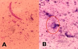
O presente estudo objetivou aprimorar a citologia fecal como método diagnóstico mais acessível e menos invasivo. Foram coletadas amostras de fezes de 14 vacas leiteiras em três momentos, com intervalos de 28 dias, obtidas por toque retal, armazenadas e transportadas em caixa térmica. A técnica consistiu na adaptação do esfregaço otológico, utilizando swab rolado sobre lâminas e duas colorações (Panótico Rápido e May-Grünwald Giemsa). A adaptação mostrou-se funcional, desde que os esfregaços fossem finos, com melhor visualização no Panótico Rápido. A técnica pode ser aplicada em contextos com recursos limitados, embora a interpretação exija experiência devido à presença de detritos fecais. Destaca-se o potencial da citologia fecal como ferramenta de triagem na medicina veterinária e a necessidade de estudos adicionais para seu refinamento e validação.
Referências
Arns, E. M. G. C., & Reis, S. T. J. (2011). Medicina veterinária legal como especialidade médico veterinária. Orientações ao médico veterinário: Manual de direitos e deveres (Vol. 1). SINDIVET.
BRASIL. Ministério da Saúde. Manual de Diagnóstico Laboratorial da Malária. Secretaria de Vigilância em Saúde. 2ª ed. Brasília, 2009. 65 p. Disponível em: https://bvsms.saude.gov.br/bvs/publicacoes/manual_diagnostico_laboratorial_malaria.pdf.
BRASIL. Ministério da Saúde. MEDEIROS, Sérgio raposo; et al. Nutrição de bovinos de corte: fundamentos e aplicações. Brasília: Embrapa, 2015. 176 p. Disponível em: https://www.embrapa.br/busca-de-publicacoes/-/publicacao/1041926/nutricao-de-bovinos-de-corte-fundamentos-e-aplicacoes.
Bugrov, N., Rudenko, P., Lutsay, V., Gurina, R., Zharov, A., Khairova, N., Molchanova, M., Krotova, E., Shopinskaya, M., Bolshakova, M., & Popova, I. (2022). Fecal Microbiota Analysis in Cats with Intestinal Dysbiosis of Varying Severity. Pathogens, 11(2), 234. https://doi.org/10.3390/pathogens11020234.
Chen, Y. C., Lin, H. Y., Chien, Y., Tung, Y. H., Ni, Y. H., & Gau, S. S. F. (2022). Altered gut microbiota correlates with behavioral problems but not gastrointestinal symptoms in individuals with autism. Brain, Behavior, and Immunity, 106, 161–178. https://doi.org/10.1016/j.bbi.2022.08.015.
Cowell, R. L., & Tyler, R. D. (2009). Diagnostic cytology and hematology of the horse E-Book. Elsevier Health Sciences.
Fallis, A. G. (2013). May-Grunwald-Giemsa staining. Journal of Chemical Information and Modeling, 53(9).
Gomez, D. E., Arroyo, L. G., Costa, M. C., Viel, L., & Weese, J. S. (2017). Characterization of the Fecal Bacterial Microbiota of Healthy and Diarrheic Dairy Calves. Journal of Veterinary Internal Medicine, 31(3), 928–939. https://doi.org/10.1111/jvim.14695.
Greene, C. E., & Appel, M. J. (2011). Canine distemper. In C. E. Greene (Ed.), Enfermedades infecciosas: Perros y gatos (pp. 25–41). Editora Interamericana.
Gresse, R., Chaucheyras-Durand, F., Fleury, M. A., Van de Wiele, T., Forano, E., & Blanquet-Diot, S. (2017). Gut microbiota dysbiosis in animals: Impact on health and disease. Frontiers in Microbiology, 8, 1–17.
Guard, B. C., Mila, H., Steiner, J. M., Mariani, C., Suchodolski, J. S., & Chastant-Maillard, S. (2017). Characterization of the fecal microbiome during neonatal and early pediatric development in puppies. PLoS ONE, 12(4), e0175718. https://doi.org/10.1371/journal.pone.0175718.
Jha, R., & Berrocoso, J. F. D. (2015). Dietary fiber and its impact on gut health and performance of monogastric animals: A review. Animal Feed Science and Technology, 212, 1–17.
Jing, Y., Yu, Y., Bai, F., Wang, L., Yang, D., Zhang, C., Qin, C., Yang, M., Zhang, D., Zhu, Y., Li, J., & Chen, Z. (2021). Effect of fecal microbiota transplantation on neurological restoration in a spinal cord injury mouse model: involvement of brain-gut axis. Microbiome, 9(1), 1–14. https://doi.org/10.1186/s40168-021-01007-y.
Johnson, K. A., Watson, A. D. J., Ettinger, S. J., & Feldman, E. C. (2004). Tratado de Medicina Interna Veterinária: Doenças do cão e do gato. Manole Ltda.
Laborclin. (2011). Manual para Antibiograma. Laborclin.
Linnehan, B. K., Kodera, S. M., Allard, S. M., Brodie, E. C., Allaband, C., Knight, R., Lutz, H. L., Carroll, M. C., Meegan, J. M., Jensen, E. D., & Gilbert, J. A. (2024). Evaluation of the safety and efficacy of fecal microbiota transplantations in bottlenose dolphins (Tursiops truncatus) using metagenomic sequencing. Journal of Applied Microbiology, 135(2). https://doi.org/10.1093/jambio/lxae026
Liu, S., Yang, L., Zhang, Y., Chen, H., Li, X., Xu, Z., Du, R., Li, X., Ma, J., & Liu, D. (2024). Review of yeast culture concerning the interactions between gut microbiota and young ruminant animals. Frontiers in Veterinary Science, 11. https://doi.org/10.3389/fvets.2024.1335765.
Munch, S. L., Nielsen, S. S., Krogh, M. A., & Capion, N. (2020). Evaluation of two fecal occult blood tests for detecting non-perforating abomasal lesions in cattle. Animals, 10(12). https://doi.org/10.3390/ani10122356.
Panziera, W., Bianchi, R. M., Galiza, G. J. N., Pereira, P. R., Mazaro, R. D., Barros, C. S. L., Kommers, G. D., Irigoyen, L. F., & Fighera, R. A. (2014). Aspectos epidemiológicos, clínicos e anatomopatológicos do linfoma em bovinos: 128 casos (1965-2013). Pesquisa Veterinária Brasileira, 34(9), 856–864. https://doi.org/10.1590/s0100-736x2014000900009.
Raskin, R. E., & Meyer, D. J. (2011). Citologia de cães e gatos: Atlas colorido e guia de interpretação. Elsevier Brasil.
R-Core-Team. (2016). R: A language and environment for statistical computing. R. Foundation for Satatical Computing.
Ryan, L., Wong, Y. T., Dwyer, K. M., Clarke, D., Kyprian, L., & Craig, J. M. (2021). Coprocytobiology: A Technical Review of Cytological Colorectal Cancer Screening in Fecal Samples. SLAS Technology, 26(6), 591–604. https://doi.org/10.1177/24726303211024562.
Sender, R., Fuchs, S., & Milo, R. (2021). Revised estimates for the number of human and bacteria cells in the body. Cell, 164, 337–340.
Sharma, A. N., Chaudhary, P., Kumar, S., Grover, C. R., & Mondal, G. (2023). Effect of symbiotic on growth performance, gut health, and immunity status in pre-ruminant buffalo calves. Scientific Reports, 13(1), 10184. https://pubmed.ncbi.nlm.nih.gov/37349514/
Trindade, D. B. M., Cavalheiro, A. L. B., Lopes, A. A., Santurio, S. T., Mazzanti, J. M., & Eduardo Schossler, J. E. (2008). Diagnóstico de criptococose canina pela citologia aspirativa por agulha fina. Ciência Rural, 38(3), 1–6.
Ueno, H., & Gonçalves, P. C. (1998). Manual para diagnóstico das helmintoses de ruminantes (Vol. 1). Japan International Cooperation Agency.
Valenciano, A. C., & Cowell, R. L. (2019). Cowell and Tyler’s diagnostic cytology and hematology of the dog and cat-E-Book. Elsevier Health Sciences. https://doi.org/DOI: 10.1016/c2016-0-02017-x.
Wolf, P. L., Ferguson, P., & Mills, I. T. (1973). Practical clinical hematology. Interpretations and Techniques. Wiley, New York, (468 p.) $8.50, 375–376. https://doi.org/10.1136/jcp.27.5.433-a.
Wu, Y., Chen, Y., Chang, Y., Zhang, X., Li, D., Wang, L., Zheng, S., Wang, R., Zhang, S., Li, J., & Zhang, L. (2020). Genotyping and identification of Cryptosporidium spp., Giardia duodenalis and Enterocytozoon bieneusi from free–range Tibetan yellow cattle and cattle–yak in Tibet, China. Acta Tropica, 212, 105671. https://doi.org/10.1016/j.actatropica.2020.105671.
Downloads
Publicado
Edição
Seção
Licença
Direitos autorais (c) 2025 Aline de Assis Rosa, Anamaia Demutris Miranda, Andressa Oliveira Machado, Maria Eduarda dos Santos, Pricilla Pozzatti, Nathan da Rocha Neves Cruz

Este trabalho está licenciado sob uma licença Creative Commons Attribution 4.0 International License.
Você tem o direito de:
Compartilhar — copiar e redistribuir o material em qualquer suporte ou formato
Adaptar — remixar, transformar, e criar a partir do material para qualquer fim, mesmo que comercial.
O licenciante não pode revogar estes direitos desde que você respeite os termos da licença. De acordo com os termos seguintes:
Atribuição
— Você deve dar o crédito apropriado, prover um link para a licença e indicar se mudanças foram feitas. Você deve fazê-lo em qualquer circunstância razoável, mas de nenhuma maneira que sugira que o licenciante apoia você ou o seu uso. Sem restrições adicionais
— Você não pode aplicar termos jurídicos ou medidas de caráter tecnológico que restrinjam legalmente outros de fazerem algo que a licença permita.