First double cervical implant in a horse in Latin America
DOI:
https://doi.org/10.31533/pubvet.v19n11e1863Keywords:
ataxia, compressive cervical myelopathy, equine cervical fusion, metal implant, neurological disease, wobbler syndromeAbstract
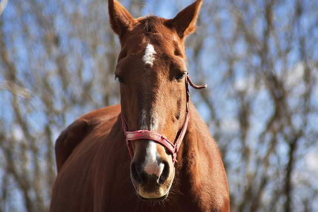
An 11-year-old Quarter Horse mare used for barrel racing, weighing 435 kg, presented with severe ataxia (grade 4). She was initially treated for pelvic and lumbar disorders and for Equine Protozoal Myeloencephalitis (EPM). However, there was no clinical improvement and no definitive diagnosis was reached. A thorough clinical and neurological examination, together with cervical radiography and contrast myelography, revealed cervical spinal cord compression at the C2–C3 and C3–C4 segments. Surgical treatment was selected, consisting of vertebral fusion (cervical arthrodesis) of the affected segments using a Brazilian metal implant. This procedure, the first double cervical fusion performed in Latin America, was successful.
References
Andrews, F. M., Matthews, H. K., & Reed, S. M. (1990). The ancillary techniques and tests for diagnosing equine neurological disease. Veterinary Medical, 85, 1325–1330.
Antonello, A. M., Pivoto, F. L., Camillo, G., Braunig, P., Sangioni, L. A., Pompermayer, E., Venturini, M. C., & Vogel, F. S. F. (2015). Investigação de anticorpos contra Sarcocystis neurona e Sarcocystis cruzi em equinos. Arquivo Brasileiro de Medicina Veterinaria e Zootecnia, 67(5). https://doi.org/10.1590/1678-4162-7374.
Bagby, G. W. (1988). Arthrodesis by the distraction-compression method using a stainless-steel implant. Orthopedics, 11(6), 931–934. https://doi.org/10.3928/0147-7447-19880601-13.
De Lahunta, A., Glass, E., & Kent, M. (2020). De Lahunta’s Veterinary neuroanatomy and clinical neurology. In de Lahunta’s Veterinary Neuroanatomy and Clinical Neurology, Fifth Edition.
Derouen, A., Spriet, M., & Aleman, M. (2016). Prevalence of anatomical variation of the sixth cervical vertebra and association with vertebral canal stenosis and articular process osteoarthritis in the horse. Veterinary Radiology and Ultrasound, 57(3), 243–258. https://doi.org/10.1111/vru.12350.
Dimock, W. W. (1950). “Wobblers” a hereditary disease in horses. Journal of Heredity, 319–323.
Dimock, W. W., & Errington, B. J. (1939). Incoordination of equidae: Wobblers. Journal of the American Veterinary Medical Association, 95, 261–267.
Dyson, S., Quiney, L., Phillips, K., Zheng, S., & Aleman, M. (2024). Radiological abnormalities of the cervicothoracic vertebrae in Warmblood horses with primary neck‐related clinical signs versus controls. Veterinary Radiology & Ultrasound, 65(6), 755–768.
Errington, B. J. (1938). Causes of wobblers. Veterinary Bulletin, 32(Sup.), 153–155.
Falco, M. J., Whitwell, K., & Palmer, A. C. (1976). An Investigation into the Genetics of ‘Wobbler Disease’ in Thoroughbred Horses in Britain. Equine Veterinary Journal, 8(4), 165–169. https://doi.org/10.1111/j.2042-3306.1976.tb03331.x.
Furr, M., Jonhson, A. L., & Reed, S. (2015). Equine Neurolgy (2nd ed.).
Glade, M. J., & Belling, T. H. (1984). Growth plate cartilage metabolism, morphology and biochemical composition in over- and underfed horses. Growth, 48(4), 473–482.
Glade, M. J., & Luba, N. K. (1987). Serum triiodothyronine and thyroxine concentrations in weanling horses fed carbohydrate by direct gastric infusion. American Journal of Veterinary Research, 48(4), 578–582. https://doi.org/10.2460/ajvr.1987.48.04.578.
Hurtig, M., Green, S. L., Dobson, H., Mikuni‐Takagaki, Y., & Choi, J. (1993). Correlative study of defective cartilage and bone growth in foals fed a low‐copper diet. Equine Veterinary Journal, 25(16 S), 66–73. https://doi.org/10.1111/j.2042-3306.1993.tb04857.x.
Knight, D. A., Gabel, A. A., Reed, S. M., Embertson, R. M., Tyznik, W. J., & Bramlage, E. R. (1985). Correlation of dietary mineral to incidence and severity of metabolic bone disease in Ohio and Kentucky. Procedings American Association of Equine Practitioners, 445–461.
Knight, D. A., Weisbrode, S. E., Schmall, L. M., Reed, S. M., Gabel, A. A., Bramlage, L. R., & Tyznik, W. I. (1990). The effects of copper supplementation on the prevalence of cartilage lesions in foals. Equine Veterinary Journal, 22(6), 445–461. https://doi.org/10.1111/j.2042-3306.1990.tb04310.x.
Kronfeld, D. S., Meacham, T. N., & Donoghue, S. (1990). Dietary aspects of developmental orthopedic disease in young horses. The Veterinary Clinics of North America. Equine Practice, 6(2), 451–465. https://doi.org/10.1016/S0749-0739(17)30551-5.
May-Davis, S. (2014). The Occurrence of a Congenital Malformation in the Sixth and Seventh Cervical Vertebrae Predominantly Observed in Thoroughbred Horses. Journal of Equine Veterinary Science, 34(11–12), 1313–1317. https://doi.org/10.1016/j.jevs.2014.09.012.
May-Davis, S., Eckelbarger, P. B., Dzingle, D., & Saber, E. (2024). Characterization and Association of the Missing Ventral Tubercle(s) from the Sixth Cervical Vertebra and Transpositions on the Ventral Surface of the Seventh Cervical Vertebra in Modern Equus ferus caballus. Animals, 14(12), 1830. https://doi.org/10.3390/ani14121830.
Mayhew, J. (2009). Tetany, tremor and postural and movement disorders. 1th International Congress of World Equine Veterinary Association.
Nixon, A. J., & Stashak, T. S. (1988). Laminectomy for relief of atlantoaxial subluxation in four horses. Journal of the American Veterinary Medicine Association, 193(6), 677–682. https://doi.org/10.2460/javma.1988.193.06.677.
Pinchbeck, G., & Murphy, D. (2001). Cervical vertebral fracture in three foals. Equine Veterinary Education, 13(1), 8–12. https://doi.org/10.1111/j.2042-3292.2001.tb01874.x.
Santinelli, I., Beccati, F., Arcelli, R., & Pepe, M. (2016). Anatomical variation of the spinous and transverse processes in the caudal cervical vertebrae and the first thoracic vertebra in horses. Equine Veterinary Journal, 48(1), 45–49. https://doi.org/10.1111/evj.12397.
Seo, J., Oh, J., Kim, P., Ju, C. Il, & Kim, S. W. (2023). Traumatic Anterior Cervical Disc Herniation Presenting as Severe Dysphagia. Diagnostics, 13(24). https://doi.org/10.3390/diagnostics13243644.
Summers, B. A., Cummings, J. F., & De Lahunta, A. (1995). Veterinary Neuropathology. Mosby-Year Book. Mosby Year Book.
Vasconcellos, L. A. S. (2025). Arthrodesis Cervical (C3C4) as a Resolution of Incoordination in Foal. 16 Th Jornadas Internacionais Hospital Muralha de Évora.
Vasconcellos, L. A. S. (1996). Artrodese Cervical em um potro. Descrição de uma Nova Técnica. XV Congresso Panamericano de Ciências Veterinárias, 121.
Vasconcellos, L. A. S. (1995). Problemas neurológicos na clínica equina. Editora Varela.
Vasconcellos, L. A. S. (2022). Neurologia e neurocirurgia equina: Princípios gerais. Editora Lusófona.
Vasconcellos, L. A. S. (2024). Irregularidade morfológica dos anéis traqueais em equinos associada ou não à malformação cervical. PUBVET, 18(5). https://doi.org/10.31533/pubvet.v18n05e1588.
Vasconcellos, L. A. S. (2025). New Brazilian implant for cervical arthrodesis in horses. Brazilian Journal of Research Environmental. https://doi.org/10.34188/bjaerv8n2-036.
Veraa, S., Bergmann, W., van den Belt, A. J., Wijnberg, I., & Back, W. (2016). Ex vivo computed tomografhic evaluations of morphology variations in equine cervical vertebrae. Veterinary Radiology and Ultrasound, 57(5). https://doi.org/10.1111/vru.12393.
Wagner, P. C., Bagby, G. W., Grant, B. D., Gallina, A., Ratzlaff, M., & Sande, R. (1979). Surgical Stabilization of the Equine Cervical Spine. Veterinary Surgery, 8(1), 85–88. https://doi.org/10.1111/j.1532-950X.1979.tb00596.x.
Watson, A. G., & Mayhew, I. G. (1986). Familial congenital occipitoatlantoaxial malformation (OAAM) in the Arabian horse. Spine, 11(4), 334–339. https://doi.org/10.1097/00007632-198605000-00007.
Downloads
Published
Issue
Section
License
Copyright (c) 2025 Luiz Alberto da Silva Vasconcellos, Beatriz Andre Marques, Caio Ferrari Teixeira, Carolina Frederico Sciacco, Cauane do Carmo Souza5, Eduarda Barreira Carvalho, Gabriel Vieira Rocha, João Vitor da Paixão Buono, Raphel Augusto da Silva Hernandez, Roberta Sargo, Thiago Escodro Dercoli

This work is licensed under a Creative Commons Attribution 4.0 International License.
Você tem o direito de:
Compartilhar — copiar e redistribuir o material em qualquer suporte ou formato
Adaptar — remixar, transformar, e criar a partir do material para qualquer fim, mesmo que comercial.
O licenciante não pode revogar estes direitos desde que você respeite os termos da licença. De acordo com os termos seguintes:
Atribuição
— Você deve dar o crédito apropriado, prover um link para a licença e indicar se mudanças foram feitas. Você deve fazê-lo em qualquer circunstância razoável, mas de nenhuma maneira que sugira que o licenciante apoia você ou o seu uso. Sem restrições adicionais
— Você não pode aplicar termos jurídicos ou medidas de caráter tecnológico que restrinjam legalmente outros de fazerem algo que a licença permita.