Renal ultrasonographic evaluation in young cats
Keywords:
Cats, early diagnosis, renal Doppler, renal resistive indexAbstract
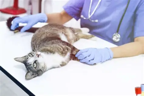
This study aimed to evaluate the morphological, qualitative, and hemodynamic ultrasonographic characteristics of the kidneys in young cats predisposed and not predisposed to congenital kidney diseases, investigating possible differences between the groups and the influence of age and body weight on the evaluated parameters. A total of 24 cats aged 3 to 12 months were examined and underwent medical history collection, physical examination, and abdominal ultrasonography using B-mode and Doppler imaging. The animals were allocated into a predisposed group, consisting of breeds at higher risk for hereditary kidney diseases, and a non-predisposed group, composed of mixed-breed cats. Most animals were in good general clinical condition and showed no evidence of renal disease. Ultrasonographic evaluation revealed that most kidneys had preserved structural characteristics, with only incidental extrarenal abnormalities observed and no distribution pattern between the groups. Comparison between the groups revealed no statistically significant differences for most of the morphometric, qualitative, and hemodynamic variables evaluated. Only the medullary thickness of the left kidney differed significantly between the groups. Analyses according to age group demonstrated a significant increase in the longitudinal length of both kidneys in older animals, whereas correlation analyses showed a positive association between age and renal cortical thickness. Body weight was positively correlated with several renal morphometric measurements but did not influence the renal resistive index. Doppler parameters remained similar between groups and age categories, suggesting stable renal perfusion during growth. It is concluded that young cats predisposed to congenital kidney diseases exhibit ultrasonographic characteristics similar to those observed in non-predisposed animals, with the main variations being associated with body growth and physiological renal maturation.
References
Ackerman, N., & Burk, R. L. (1996). Small animal radiology and ultrasonography a diagnostic atlas and text. WB Saunders Company. USA.
Avni, F. E., Garel, C., Cassart, M., D’Haene, N., Hall, M., & Riccabona, M. (2012). Imaging and classification of congenital cystic renal diseases. American Journal of Roentgenology, 198(5). https://doi.org/10.2214/AJR.11.8083.
Barthez, P. Y., Rivier, P., & Begon, D. (2003). Prevalence of polycystic kidney disease in Persian and Persian related cats in France. Journal of Feline Medicine and Surgery, 5(6), 345–347. https://doi.org/10.1016/S1098-612X(03)00052-4.
Berry, C. R., Nelson, N. C., & Winter, M. D. (2023). Atlas of Small Animal Diagnostic Imaging. In Atlas of Small Animal Diagnostic Imaging. https://doi.org/10.1002/9781119533221.
Calhau, D. S., Pellegrini, G. S., Pellin, I. C., Nagamine, K. M., Almeida, Y. T. C., & Eiras, C. E. (2024). Doença renal crônica em gatos. PUBVET, 18(2), e1551. https://doi.org/10.31533/pubvet.v18n02e1551.
Carvalho, C. F. (2018). Ultrassonografia em pequenos animais. Editora Roca.
Carvalho, C. F., Chammas, M. C., & Cerri, G. G. (2008). Princípios físicos do Doppler em ultrassonografia: Revisão bibliográfica. Ciência Rural, 38, 872–879.
Castiglioni, M. C. R., Rahal, S. C., Silva, J. P., Ichikawa, R. S., & Mamprim, M. J. (2025). Doppler ultrasound for the assessment of renal perfusion in kittens up to three months of age and young adult cats. Research in Veterinary Science, 187, 105590. https://doi.org/10.1016/j.rvsc.2025.105590.
Castiglioni, M. C. R., Rahal, S. C., Silva, J. P., & Mamprim, M. J. (2022). Comparison of ultrasonographic renal measurements in kittens up to 3 months of age and young cats. Journal of Feline Medicine and Surgery, 24(12), e465–e471. https://doi.org/10.1177/1098612X221119392.
Cavalcante, R. S. G., Alegrio, B. L., Parreira, L. M., & Goulart, I. C. (2025). Terapias gênicas para doenças neurodegenerativas hereditárias. Journal Archives of Health, 6(4). https://doi.org/10.46919/archv6n4espec-15998.
Debruyn, K., Haers, H., Combes, A., Paepe, D., Peremans, K., Vanderperren, K., & Saunders, J. H. (2012). Ultrasonography of the feline kidney. Journal of Feline Medicine and Surgery, 14(11), 794–803. https://doi.org/10.1177/1098612x12464461.
Greco, D. S. (2001). Congenital and inherited renal disease of small animals. The Veterinary Clinics of North America. Small Animal Practice, 31(2). https://doi.org/10.1016/S0195-5616(01)50211-9.
Griffin, S. (2020). Feline abdominal ultrasonography: what’s normal? what’s abnormal? The kidneys and perinephric space. Journal of Feline Medicine and Surgery, 22(5), 409–427. https://doi.org/10.1177/1098612X20917598.
JAMOVI. (2021). The Jamoviprojet, VERSION 1.6 [Broadcast].
Jericó, M. M., Andrade Neto, J. P., & Kogika, M. M. (2015). Tratado de medicina interna de cães e gatos. Roca Ltda.
Kealy, J. K., McAllister, H., & Graham, J. P. (2012). Radiologia e ultrassonografia do cão e do gato (Vol. 1, 96–109). Elselvier Saunders.
Kongtasai, T., Paepe, D., Meyer, E., Mortier, F., Marynissen, S., Stammeleer, L., Defauw, P., & Daminet, S. (2022). Renal biomarkers in cats: A review of the current status in chronic kidney disease. Journal of Veterinary Internal Medicine, 36(2). https://doi.org/10.1111/jvim.16377.
Lees, G. E. (1996). Congenital renal diseases. In The Veterinary Clinics of North America. Small Snimal Practice (Vol. 26, Number 6). https://doi.org/10.1016/S0195-5616(96)50133-6.
Lopes, M. C. T., Honorato, R. A., Sousa Gomes, M., Silva, E. M. C., Santos, A. C. C., & Albuquerque, W. R. (2015). Doença renal policística felina: Relato de caso. PUBVET, 9(3), 115–118.
Lourenço, P. Pietro, & Albuquerque, K. D. (2019). O uso da dimetilarginina simétrica (SDMA) no diagnóstico e estadiamento da doença renal crônica em felinos: Revisão de literatura. Revista de Educação Continuada Em Medicina Veterinária e Zootecnia Do CRMV-SP, 17(2), 24–33. https://doi.org/10.36440/recmvz.v17i2.37918.
Maher, M. A., Palacio, L., Henao, J. C., & Elsharkawy, S. H. (2024). Gross morphological and ultrasonographic dimensions of normal feline kidney with reference to resistive index. Scientific Reports, 14(1). https://doi.org/10.1038/s41598-024-75341-0.
Mazutti, L. C., & Ferreira, A. B. G. (2021). Doença renal crônica em gatos: A importância dos estadiamentos e do diagnóstico precoce: revisão de literatura. Arquivos Brasileiros de Medicina Veterinária, 4(1), 155–194.
Novellas, R., Gopegui, R. R., & Espada, Y. (2010). Assessment of renal vascular resistance and blood pressure in dogs and cats with renal disease. Veterinary Record, 166(20), 618–623. https://doi.org/10.1136/vr.b4820.
Oliveira, L. L., Attallah, F. A., Santos, C. L., Wakofs, T. N., Rodrigues, M. C. D., & Santos, A. E. (2005). O uso da ultrassonografia para o diagnóstico de Dioctophyma renale em cão: Relato de caso. Annals V Conferência Sul-Americana de Medicina Veterinária.
Oliveira, M. R. H., Sousa, P. F. S., Martins, G. O., Simão, E. P., & Martins, L. R. (2025). Terapias gênicas e suas aplicações em Doenças Hereditárias. Brazilian Journal of Health Review, 8(1), 10–16. https://doi.org/10.34119/bjhrv8n1-488.
Rey, M. S. (2013). Doenças renais hereditárias e congênitas felinas. Veterinary Focus, 3, 10–16.
Reznik, V. M., & Budorick, N. E. (1995). Prenatal detection of congenital renal disease. Urologic Clinics of North America, 22(1). https://doi.org/10.1016/s0094-0143(21)01013-2.
Sargent, H. J., & Elliot, J. (2020). Detection of early chronic kidney disease in cats. Veterinary Focus, 30(1), 30–38.
Scardoeli, B. (2017). Doença renal crônica em cães e gatos: revisão bibliográfica e estudo retrospectivo.
Silva, L. R., Rodrigues, G. B., Barrio, M. A. M., & Brisola, M. L. (2021). Avaliação da dimetilarginina simétrica (SDMA) como marcador de filtração glomerular em cães e gatos pré-azotêmicos. Brazilian Journal of Animal and Environmental Research, 4(1). https://doi.org/10.34188/bjaerv4n1-015.
Sim, H., An, Y., Kim, S. S., Kwon, D., Lee, J., Lee, K., & Yoon, H. (2025). Feline Renal Cortical Thickness–Aortic Diameter Ratio in Healthy Versus Diseased Kidneys: Comparative Ultrasonographic Evaluation. Veterinary Radiology and Ultrasound, 66(5). https://doi.org/10.1111/vru.70090.
Slinkard, P. T., Anson, A., Specchi, S., Frau Tascon, M., Muñiz-Moris, L., Penninck, D., & Ondreka, N. E. (2023). Imaging features of renal ectopia and fusion in 13 cats. Journal of Feline Medicine and Surgery, 25(10). https://doi.org/10.1177/1098612X231196810.
Souza Júnior, J. C., Costa, J. R., Favero, P. V. S., Leal, A. A., Miranda, H. L. A., Silva, L. O., & Novaes, V. R. F. (2025). A utilização do dimetilarginina simétrica como marcador precoce da doença renal crônica em pequenos animais: Relato de caso. PUBVET, 19(7), 1.
Tipisca, V., Murino, C., Cortese, L., Mennonna, G., Auletta, L., Vulpe, V., & Meomartino, L. (2015). Resistive index for kidney evaluation in normal and diseased cats. Journal of Feline Medicine and Surgery, 18(6), 471–475. https://doi.org/10.1177/1098612X15587573.
Tokas, T., Herrmann, T. R. W., Skolarikos, A., & Nagele, U. (2019). Pressure matters: intrarenal pressures during normal and pathological conditions, and impact of increased values to renal physiology. World Journal of Urology, 37(1), 125–131.
Xavier Júnior, F. A. F., Morais, G. B., Dutra, M. S., Freitas, M. M., Araujo, S. L., Araújo, D. V., & Evangelista, J. S. A. M. (2019). Doença Renal Aguda em gatos: Conquistas e desafios. Medicina Veterinária (UFRPE), 13(3). https://doi.org/10.26605/medvet-v13n3-3308.
Downloads
Published
Issue
Section
License
Copyright (c) 2026 Eduarda Cardoso de Miranda, Victoria Cristina dos Santos, Ketlyn Naiara Kolb, Lidiane Sievert, Matheus Candano de Brito, Matheus Folgearini Silveira, Carlize Lopes, Raquel de Souza Lemos de Oliveira

This work is licensed under a Creative Commons Attribution 4.0 International License.
Você tem o direito de:
Compartilhar — copiar e redistribuir o material em qualquer suporte ou formato
Adaptar — remixar, transformar, e criar a partir do material para qualquer fim, mesmo que comercial.
O licenciante não pode revogar estes direitos desde que você respeite os termos da licença. De acordo com os termos seguintes:
Atribuição
— Você deve dar o crédito apropriado, prover um link para a licença e indicar se mudanças foram feitas. Você deve fazê-lo em qualquer circunstância razoável, mas de nenhuma maneira que sugira que o licenciante apoia você ou o seu uso. Sem restrições adicionais
— Você não pode aplicar termos jurídicos ou medidas de caráter tecnológico que restrinjam legalmente outros de fazerem algo que a licença permita.