Detection and morphological classification of neutrophils using convolutional neural networks to support left shift diagnosis
DOI:
https://doi.org/10.31533/pubvet.v20n5e1959Keywords:
band neutrophils, left shift, veterinary hematology, neutrophils, deep learning, yolov8Abstract
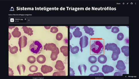
A left shift is characterized by an increased number of immature neutrophils (band cells) in the bloodstream, indicating a bone marrow response to acute infection or inflammation. Leukocyte counting and the morphological differentiation between segmented and band neutrophils are crucial steps in laboratory screening. Although automated hematology analyzers are available, they are expensive and often face challenges in accurately distinguishing cell morphology, particularly in three-part differential (3-DIFF) systems. This limitation frequently generates system alerts (flags), requiring manual microscopic review to complement the analysis. However, manual counting is time-consuming and subject to interobserver variability. This study aimed to evaluate the feasibility of the YOLOv8 computer vision architecture for the detection and classification of neutrophils. A dataset of digital microscopic images obtained from the Kaggle platform was curated and annotated using Roboflow software, with the application of data augmentation techniques. The model was trained in a cloud-computing environment using Google Colab. The results demonstrated promising algorithm performance, showing technical feasibility for both cell localization and differentiation between band and segmented neutrophils, achieving a Mean Average Precision (mAP50) of 0.90. Furthermore, a web-based application prototype was developed as a proof of concept to demonstrate the model's inference capabilities. These findings suggest that Deep Learning technology has significant potential for automated cellular differentiation. In future developments, its direct integration into microscopy systems may serve as a valuable tool for supporting and standardizing left shift diagnosis.
References
Asghar, R., Kumar, S., Shaukat, A., & Hynds, P. (2024). Classification of white blood cells (leucocytes) from blood smear imagery using machine and deep learning models: A global scoping review. PLoS ONE, 19(6), e0292026. https://doi.org/10.1371/journal.pone.0292026.
Baruah, D. K., Boruah, K., Barman, N. N., Deka, A., Bharali, A., & Buragohain, L. (2025). Automated Detection of Canine Babesia Parasite in Blood Smear Images Using Deep Learning and Contrastive Learning Techniques. Parasitologia, 5(2). https://doi.org/10.3390/parasitologia5020023.
Black, F. S., Melo, A. J. P., Giordani, T. S., Cripa, F. B., Scapin, D., & Machado, L. P. (2025). Estudo Comparativo da Contagem Diferencial de Leucócitos por Microscopia e por Analisador Hematológico Automático. Revista Ibero-Americana de Humanidades, Ciências e Educação, 11(3), 2164–2174. https://doi.org/10.51891/rease.v11i3.18545.
Borges, L. F., & Siqueira, L. O. (2009). Validação de tecnologia 5diff do analisador hematológico sysmex XS-1000i para laboratório de pequeno/médio porte. Revista Brasileira de Hematologia e Hemoterapia, 31(4), 231–236. https://doi.org/10.1590/s1516-84842009005000059.
Eurell, J. A., & Frappier, B. L. (2012). Histologia Veterinária de Dellmann (Vol. 1). Guanabara Koogan.
Ferreira, Â. M., Castro, M. L. R., Murador, P., Ramos, R. J., & Castilho, S. L. (2014). Imuno-hematologia laboratorial. In Ministério da Saúde (Vol. 1).
González-Recio, O., Maturana, E. L., Vega, A. T., Engelman, C. D., & Broman, K. W. (2009). Detecting single-nucleotide polymorphism by single-nucleotide polymorphism interactions in rheumatoid arthritis using a two-step approach with machine learning and a Bayesian threshold least absolute shrinkage and selection operator (LASSO) model. BMC Proceedings, 3(7), S63.
Makrai, L., Fodróczy, B., Nagy, S. Á., Czeiszing, P., Csabai, I., Szita, G., & Solymosi, N. (2023). Annotated dataset for deep-learning-based bacterial colony detection. Scientific Data, 10(1). https://doi.org/10.1038/s41597-023-02404-8.
Paes, P. R. O., Leme, F. O. P., & Carneiro, R. A. (2009). Hematologia dos animais domésticos. FEPMVZ, Caderno Didático.
Roboflow. (2025). Roboflow: Computer Vision Platform. Roboflow Inc., Des Moines, IA, USA. Disponível em: https://roboflow.com.
Souza, E. P., Gomes, C. M., Barroso, D. H., Miranda, V. L., & Gurgel-Gonçalves, R. (2019). Aplicações do Deep Learning para diagnóstico de doenças e identificação de insetos vetores. Saúde em Debate, 43(spe2), 147–154. https://doi.org/10.1590/0103-11042019s211.
Thrall, M. A., Weiser, G., Allison, R. W., & Campbell, T. W. (2022). Veterinary hematology, clinical chemistry, and cytology. John Wiley & Sons.
Downloads
Published
Issue
Section
License
Copyright (c) 2026 Luan Carlos Verteiro Pereira, Vanessa Aparecida Feijó de Souza

This work is licensed under a Creative Commons Attribution 4.0 International License.
Você tem o direito de:
Compartilhar — copiar e redistribuir o material em qualquer suporte ou formato
Adaptar — remixar, transformar, e criar a partir do material para qualquer fim, mesmo que comercial.
O licenciante não pode revogar estes direitos desde que você respeite os termos da licença. De acordo com os termos seguintes:
Atribuição
— Você deve dar o crédito apropriado, prover um link para a licença e indicar se mudanças foram feitas. Você deve fazê-lo em qualquer circunstância razoável, mas de nenhuma maneira que sugira que o licenciante apoia você ou o seu uso. Sem restrições adicionais
— Você não pode aplicar termos jurídicos ou medidas de caráter tecnológico que restrinjam legalmente outros de fazerem algo que a licença permita.