Pulmonary artery dilation secondary to pulmonary hypertension in dog
Case report
DOI:
https://doi.org/10.31533/pubvet.v20n3e1928Keywords:
Dog, echocardiography, right-sided heart failureAbstract
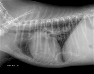
Pulmonary hypertension (PH) occurs in a variety of diseases and is characterized by increased pulmonary vascular pressure. This alteration may lead to hemodynamic overload of the pulmonary artery, right ventricle, and right atrium. As a consequence, cardiac remodeling and progressive structural damage may develop. This condition may be primary or, more commonly, secondary to cardiac, respiratory, or systemic diseases, presenting with nonspecific clinical signs that are generally related to the underlying disorder. The present study aims to report and discuss the case of a five-year-old male dog diagnosed with PH possibly associated with respiratory disease, without etiological confirmation. The animal was presented with pulmonary artery dilation and signs of right-sided heart failure, including ascites, jugular vein distension, occasional coughing, respiratory effort, and lethargy. The diagnosis was established based on clinical signs, physical examination, thoracic radiography, and echocardiography. The underlying cause was not identified, as the patient rapidly progressed to death due to cardiac arrest. This report highlights the importance of monitoring animals affected by PH and, whenever possible, identifying the underlying etiology in order to implement appropriate treatment, reduce the risk of complications, improve prognosis, and provide better quality of life for the patient.
References
Aleixo, M. F. M. (2024). Hipertensão pulmonar em cães. Escola Veterinária Vasco da Gama.
Braz, J. B., Camacho, A. A., Canola, R. A. M., Beluque, T., Zacché, E., Silva-Filho, J. C., Zamora, W. A., & Sousa, M. G. (2021). Echocardiographic features of pre- and postcapillary pulmonary hypertension with a focus on the right ventricle in dogs. Pesquisa Veterinária Brasileira, 41, e6810. https://doi.org/10.1590/1678-5150-PVB-6810.
Caetano, I. R., Costa, B. G., & Paulino Júnior, D. (2023). Erlichiose monocítica canina: Atualidades sobre a doença. Revista Ibero-Americana de Humanidades, Ciências e Educação, 9(9). https://doi.org/10.51891/rease.v9i9.11415.
Feldhütter, E. K., Domenech, O., Vezzosi, T., Tognetti, R., Eberhard, J., Friederich, J., & Wess, G. (2022). Right ventricular size and function evaluated by various echocardiographic indices in dogs with pulmonary hypertension. Journal of Veterinary Internal Medicine, 36(6), 1882–1891. https://doi.org/10.1111/jvim.16496.
Nazzareno, G., Hoeper, M. M., Humbert, M., Torbicki, A., Vachiery, J. L., Barbera, J. A., Beghetti, M., Ru, P. C., & Simoneau, G. (2009). Orientações para o diagnóstico e tratamento da hipertensão pulmonar. European Heart Journal, 30, 2493–2537. https://doi.org/10.1016/j.repc.2011.12.002.
Reinero, C., Visser, L. C., Kellihan, H. B., Masseau, I., Rozanski, E., Clercx, C., Williams, K., Abbott, J., Borgarelli, M., & Scansen, B. A. (2020). ACVIM consensus statement guidelines for the diagnosis, classification, treatment, and monitoring of pulmonary hypertension in dogs. Journal of Veterinary Internal Medicine, 34(2), 549–573. https://doi.org/10.1111/jvim.15725.
Yuchi, Y., Suzuki, R., Saito, T., Yasumura, Y., Teshima, T., Matsumoto, H., & Koyama, H. (2023). Echocardiographic characteristics of dogs with pulmonary hypertension secondary to respiratory diseases. Journal of Veterinary Internal Medicine, 37(5), 1656–1666. https://doi.org/10.1111/jvim.16836.
Downloads
Published
Issue
Section
License
Copyright (c) 2026 Aline Hochstetler de Mesquita, Gustavo Cavinato Herrera

This work is licensed under a Creative Commons Attribution 4.0 International License.
Você tem o direito de:
Compartilhar — copiar e redistribuir o material em qualquer suporte ou formato
Adaptar — remixar, transformar, e criar a partir do material para qualquer fim, mesmo que comercial.
O licenciante não pode revogar estes direitos desde que você respeite os termos da licença. De acordo com os termos seguintes:
Atribuição
— Você deve dar o crédito apropriado, prover um link para a licença e indicar se mudanças foram feitas. Você deve fazê-lo em qualquer circunstância razoável, mas de nenhuma maneira que sugira que o licenciante apoia você ou o seu uso. Sem restrições adicionais
— Você não pode aplicar termos jurídicos ou medidas de caráter tecnológico que restrinjam legalmente outros de fazerem algo que a licença permita.